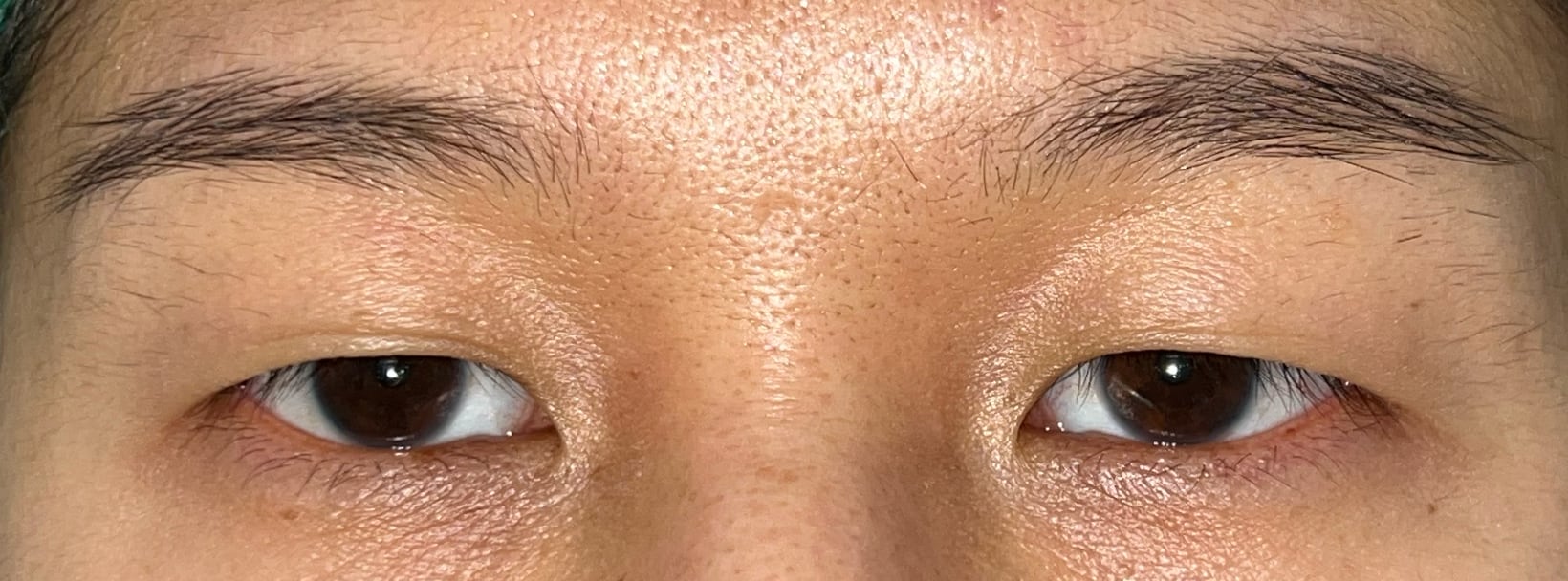
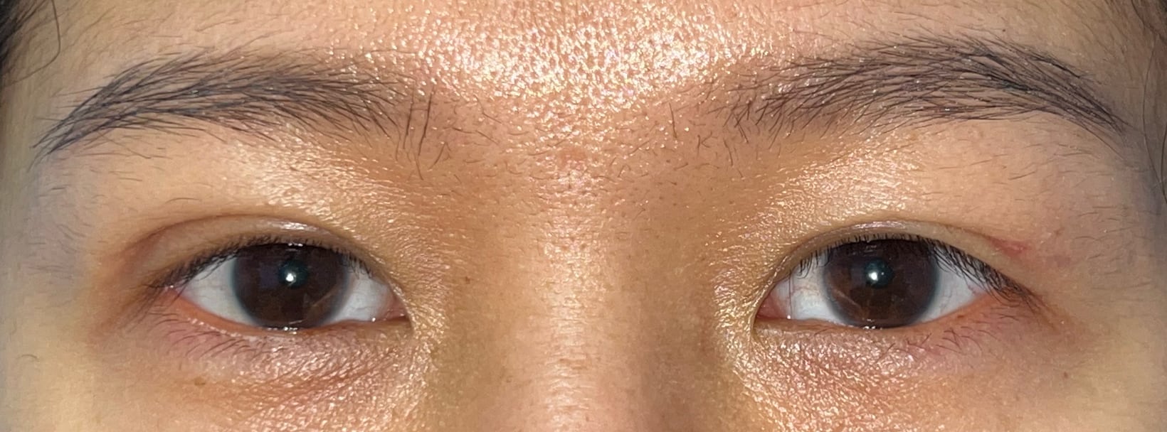
Upper Blepharoplasty
Upper eyelid result shown at four months after surgery.
Before and after photographs are shown together with the procedure and follow-up period to help explain what changed in each case. Results vary between patients and should not be used to predict an individual outcome.
Upper eyelid result shown at four months after surgery.
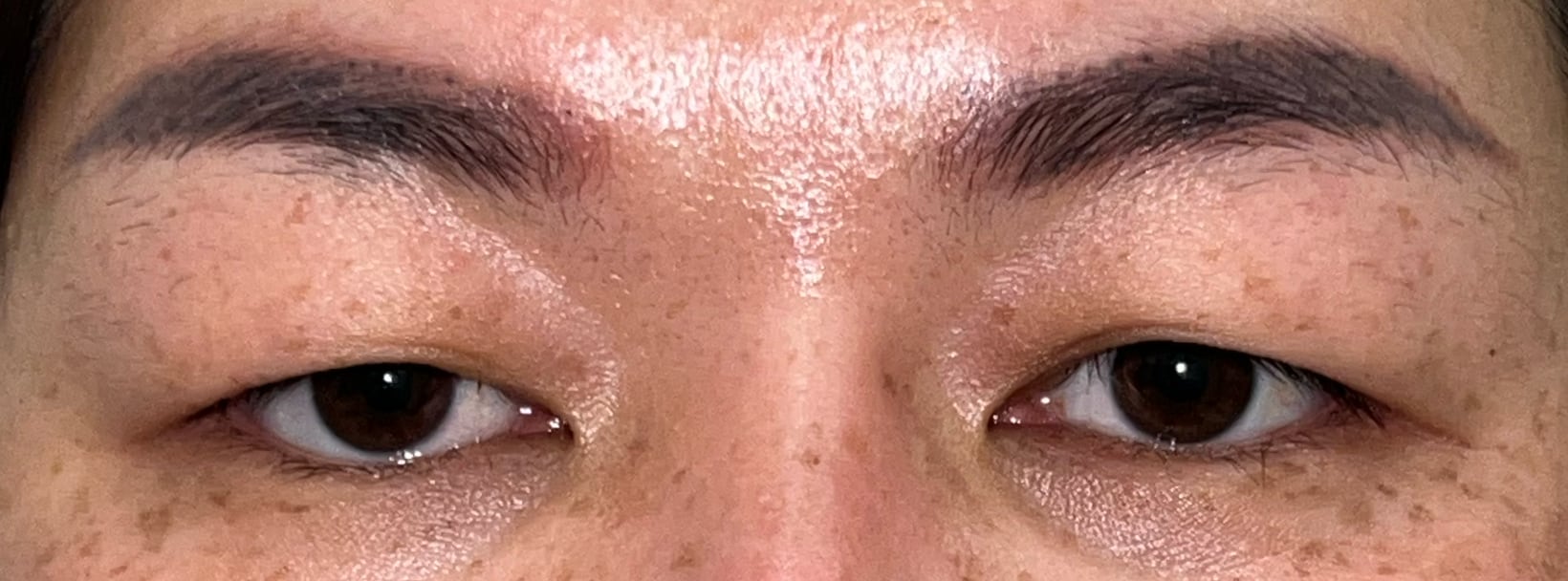
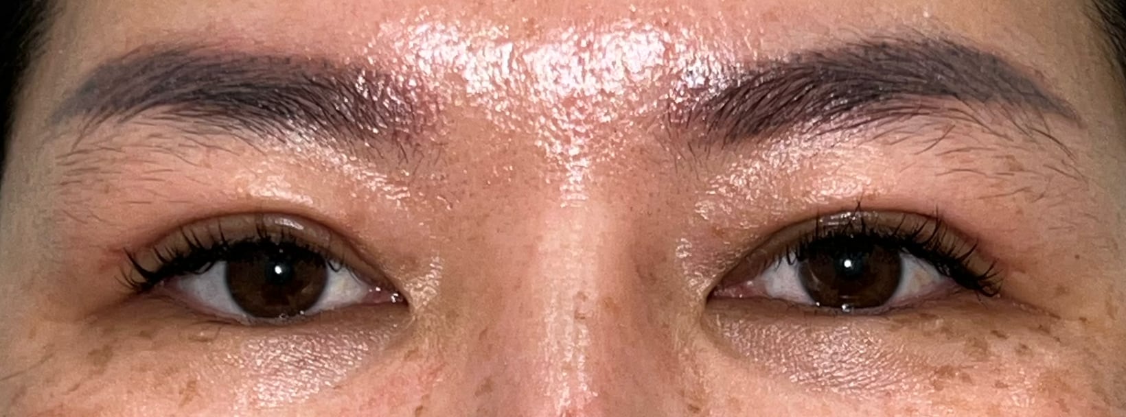
Upper eyelid result shown at two months after surgery.
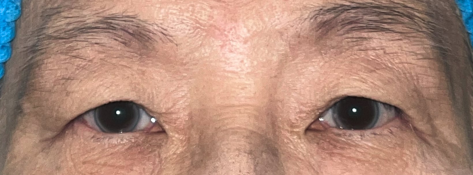
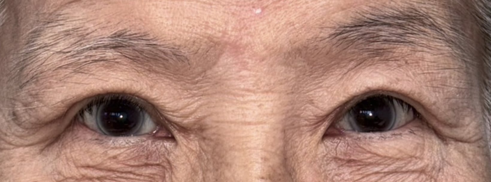
Upper eyelid result shown at three months after surgery.
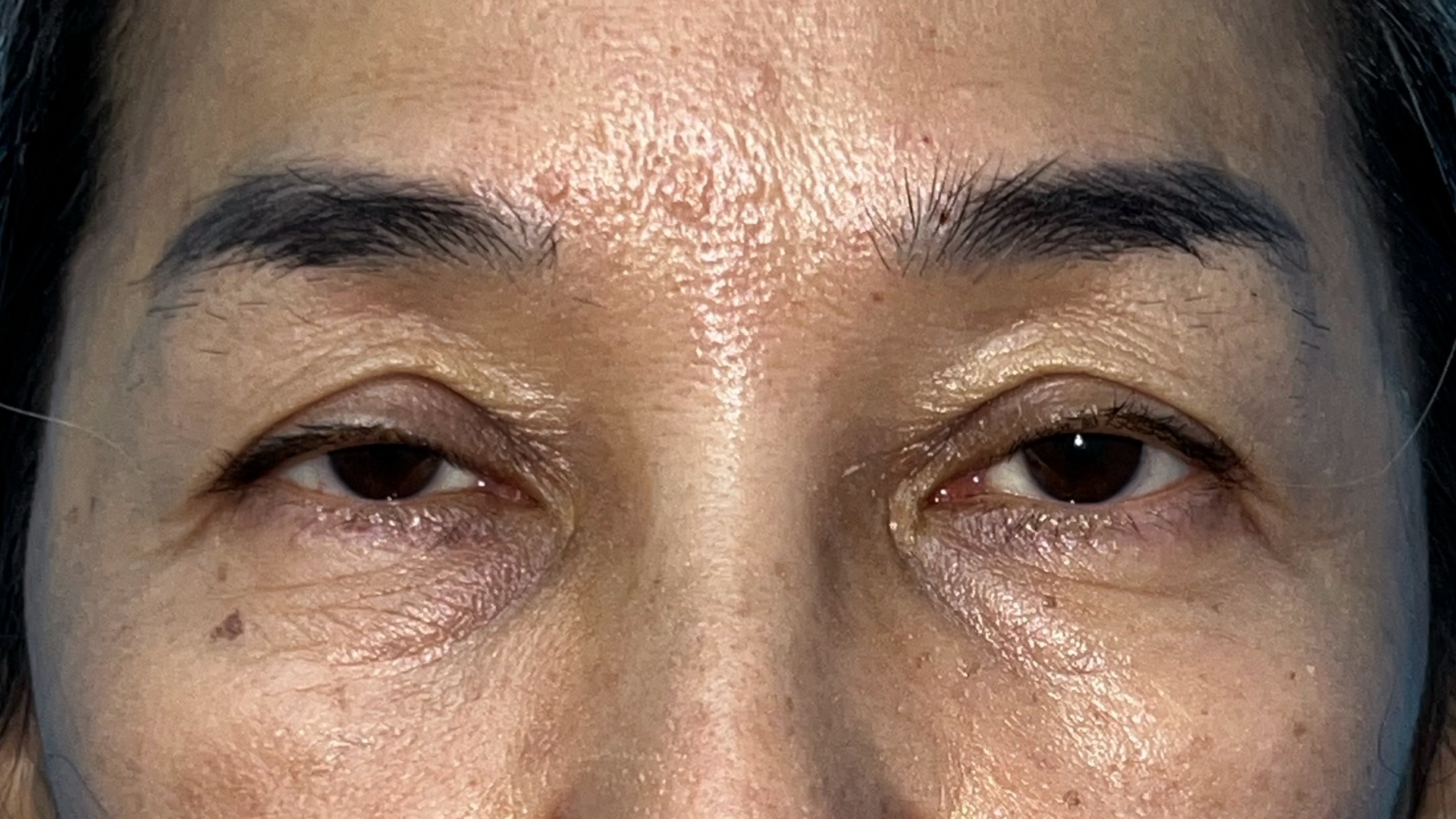
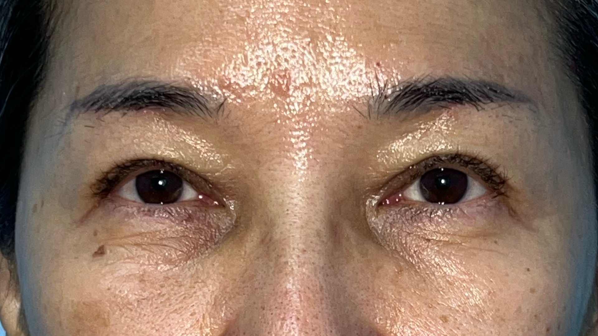
Ptosis surgery result shown at six months after levator advancement.
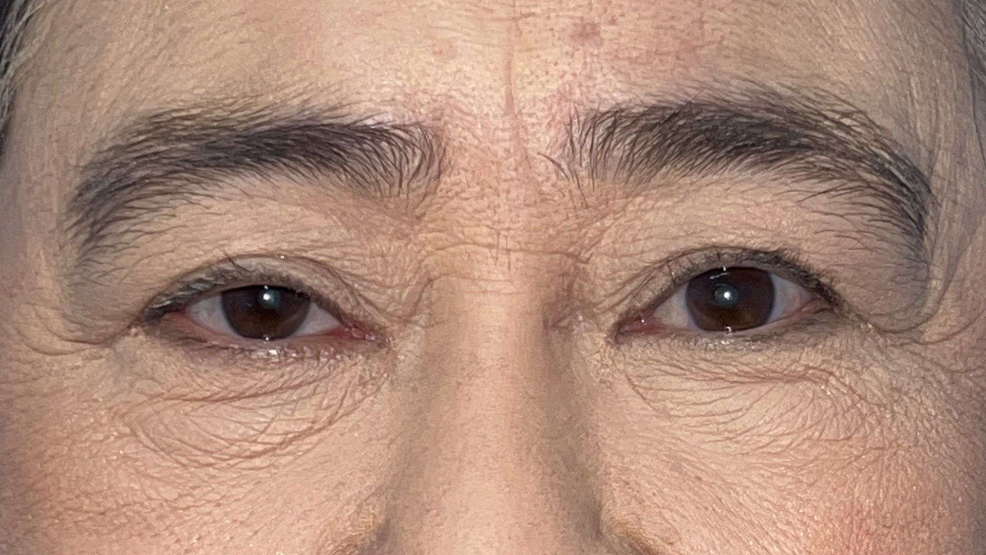
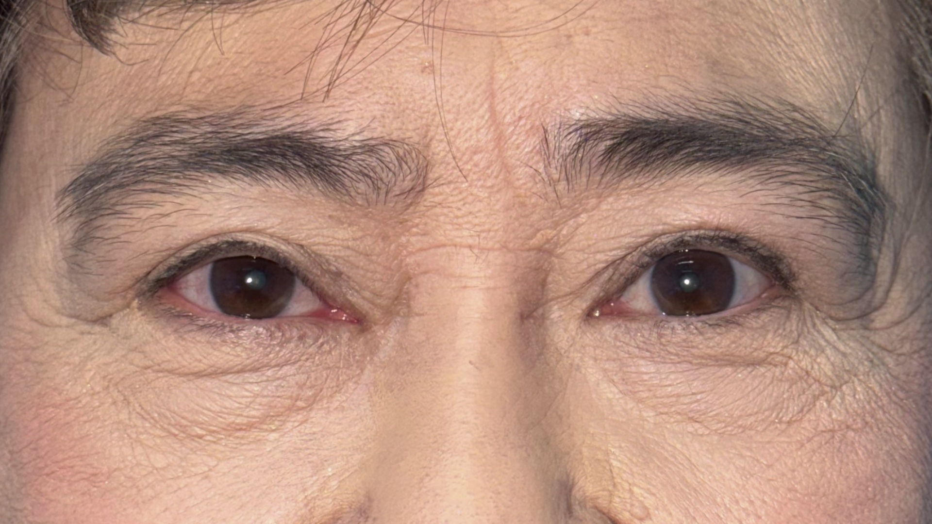
Ptosis surgery result shown at eight months after levator advancement.
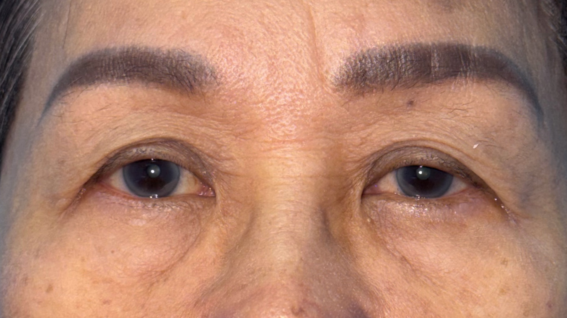
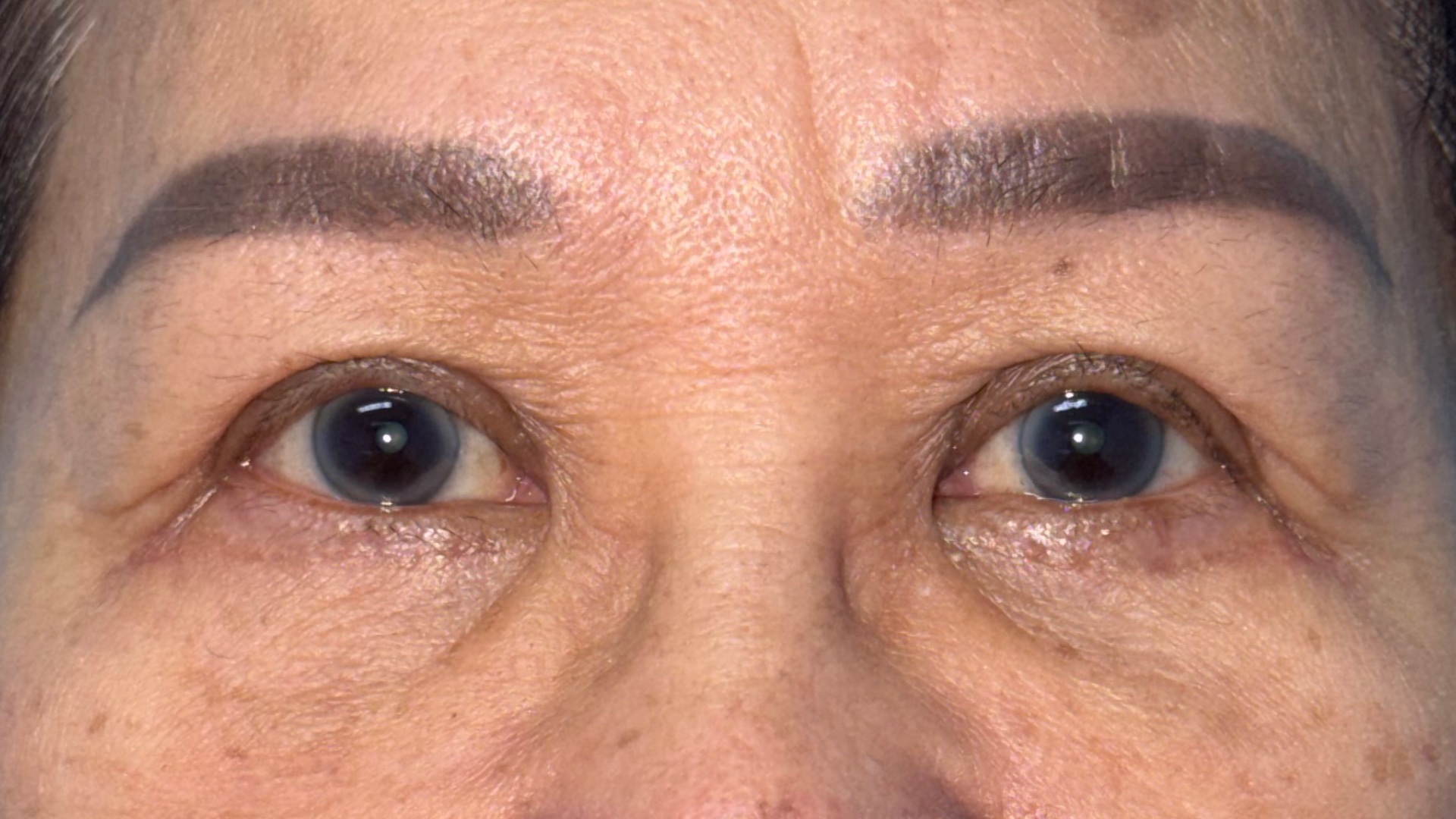
Ptosis surgery result shown at 4.5 months after levator advancement.
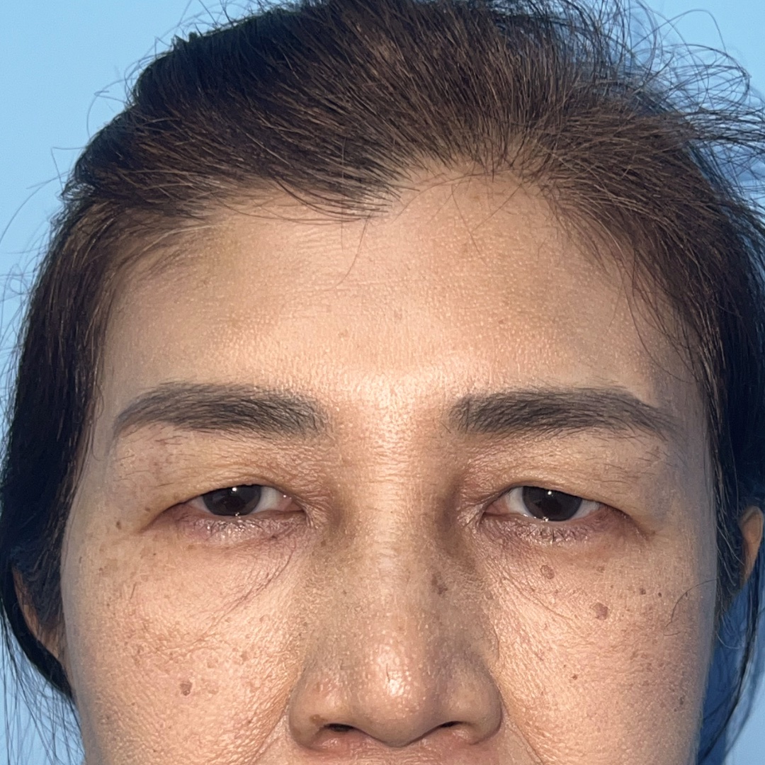
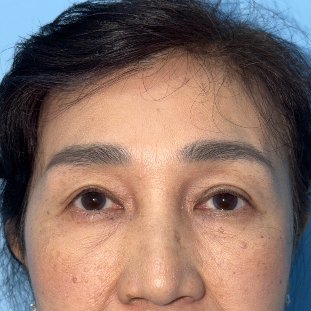
Significant brow ptosis with coexisting levator dysfunction. The treatment plan addressed the brow descent and eyelid ptosis as separate anatomical problems.
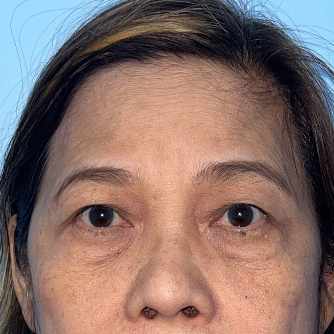
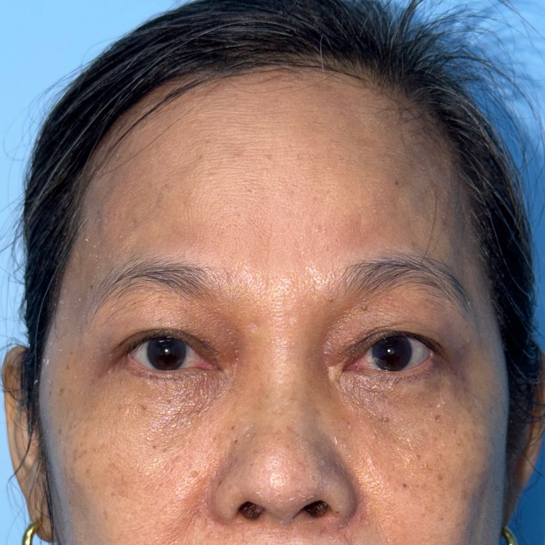
Combined surgery addressing brow descent together with lower eyelid concerns. The procedures were planned as separate anatomical components within the same treatment.
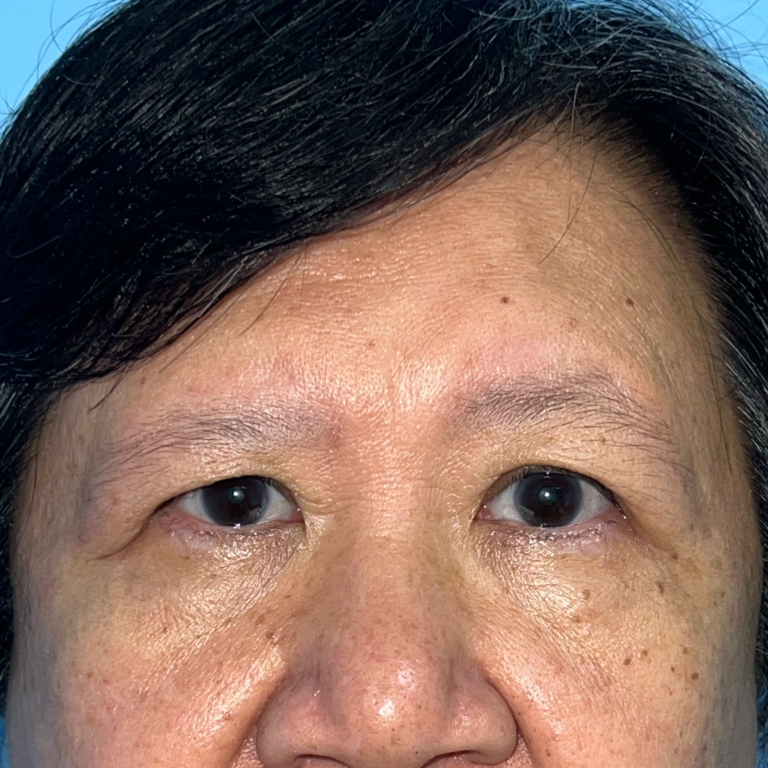
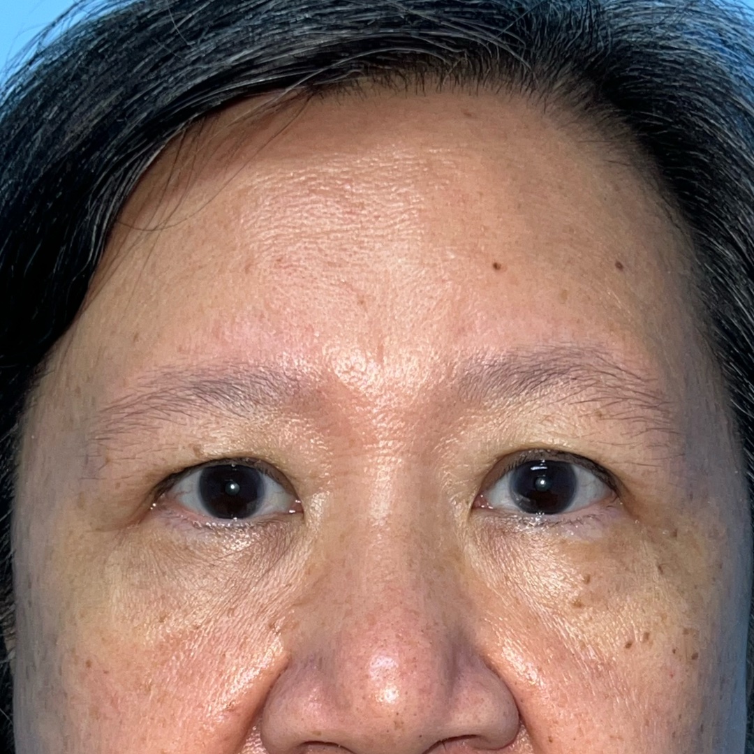
Endoscopic brow lift result shown at 2 months, with brow position and surrounding tissues still continuing to settle.
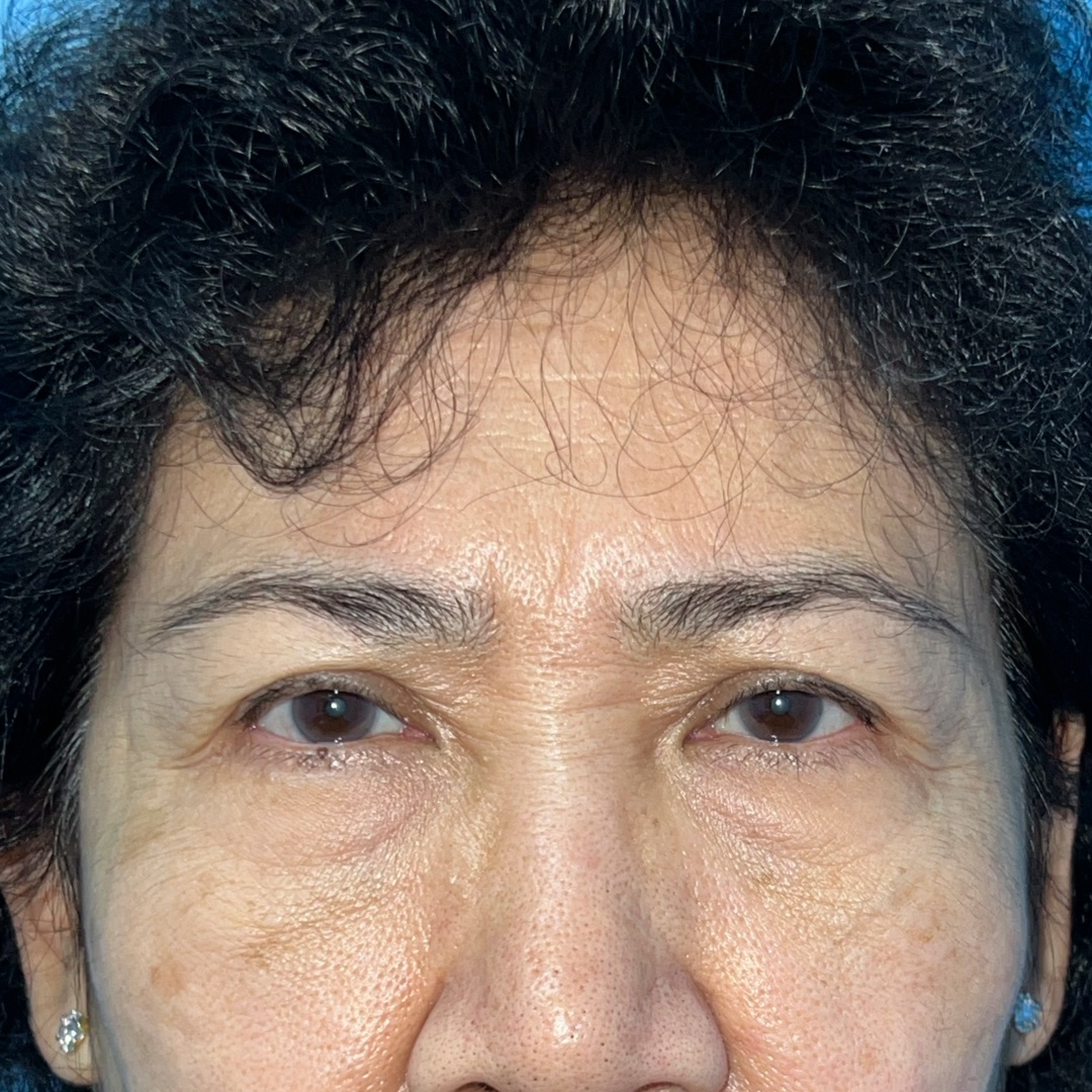
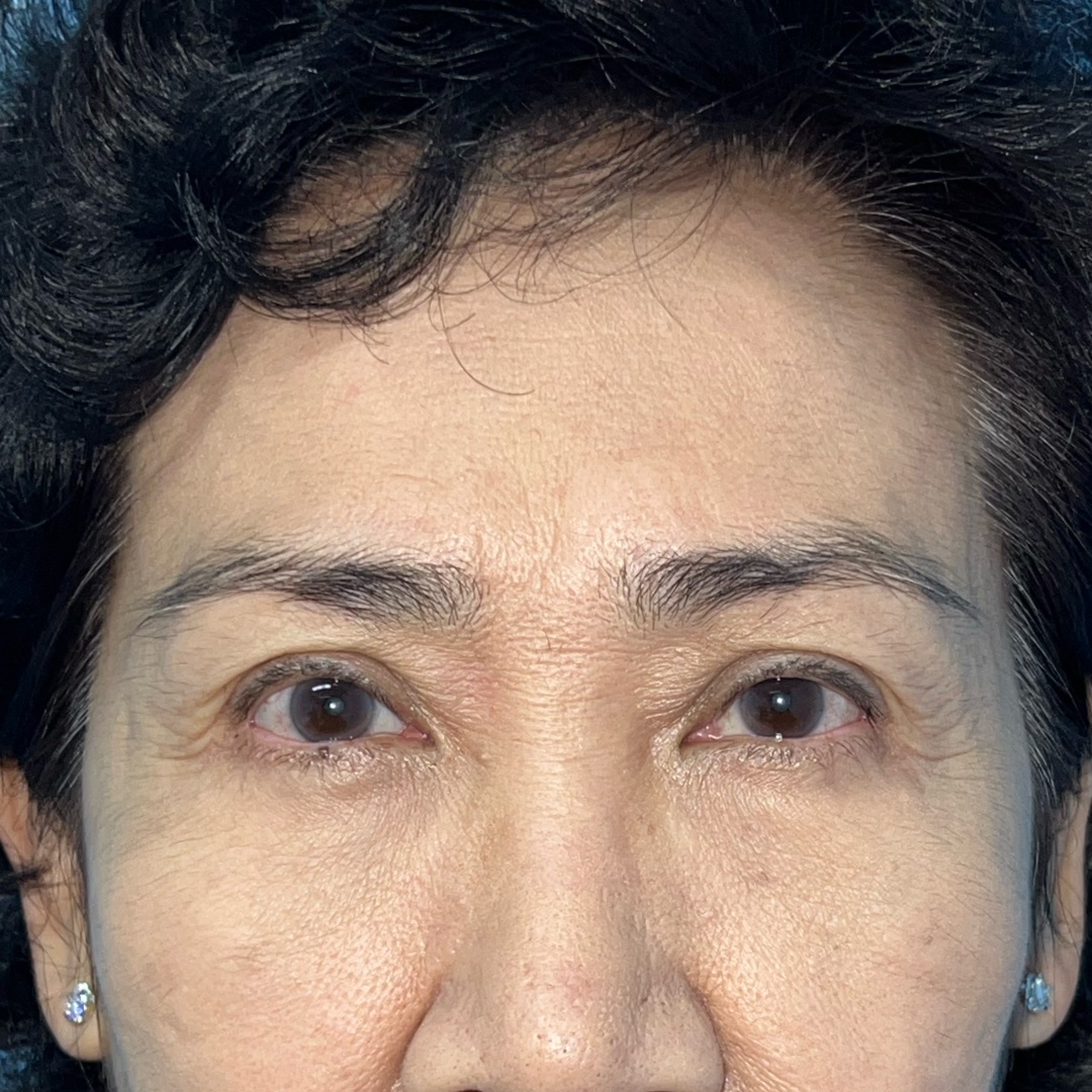
Combined surgery addressing brow descent together with lower eyelid concerns, shown at 10 months after surgery.
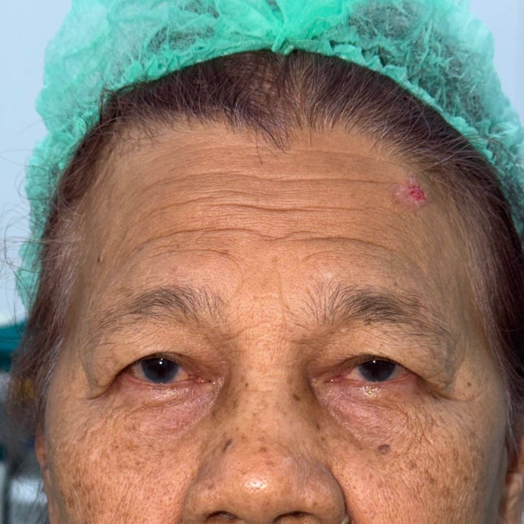
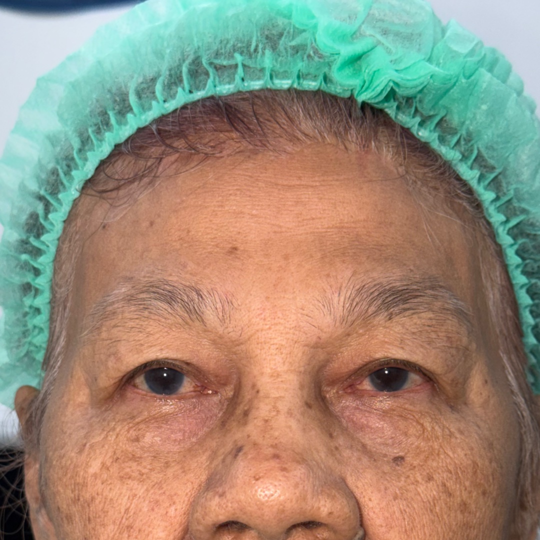
Hairline forehead lift result shown at 5 months after surgery.
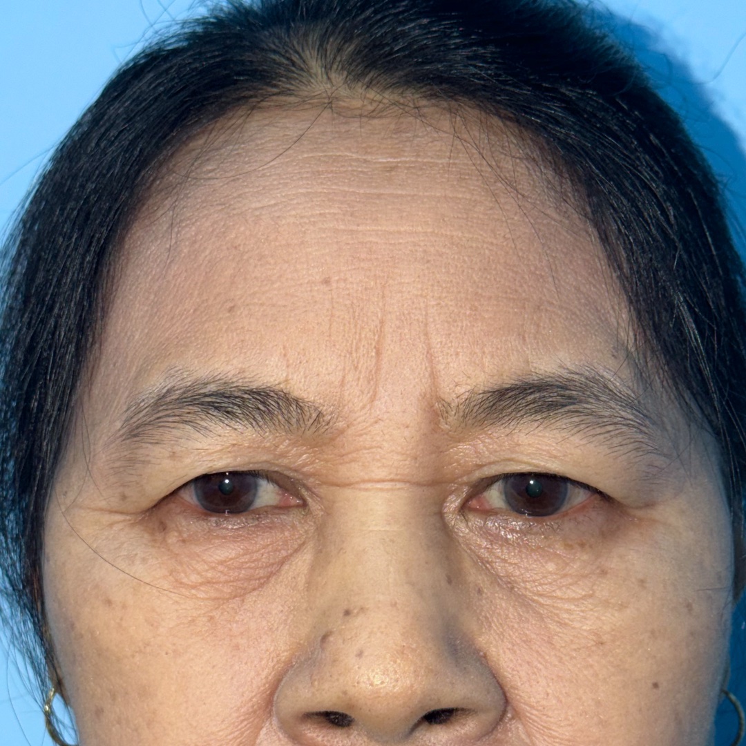
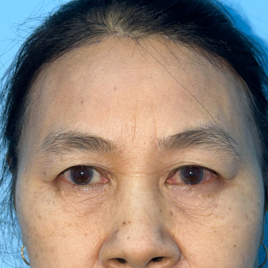
Hairline forehead lift result shown at 3 months after surgery.
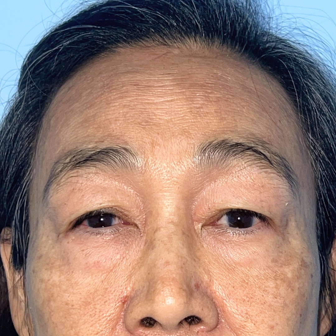
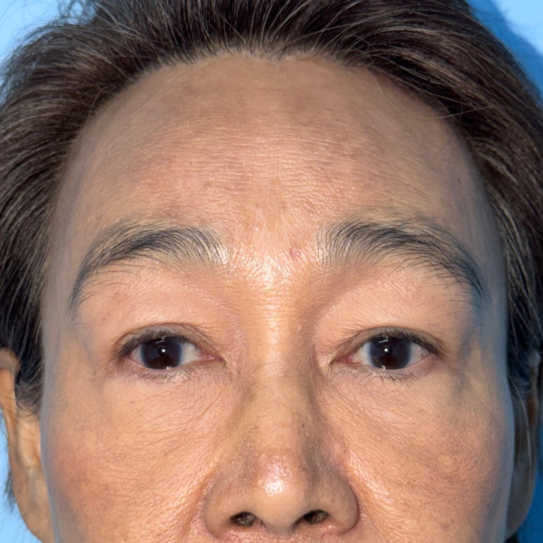
Brow ptosis with excess upper eyelid skin. A hairline approach was selected in the setting of prominent forehead wrinkles and a relatively high forehead.
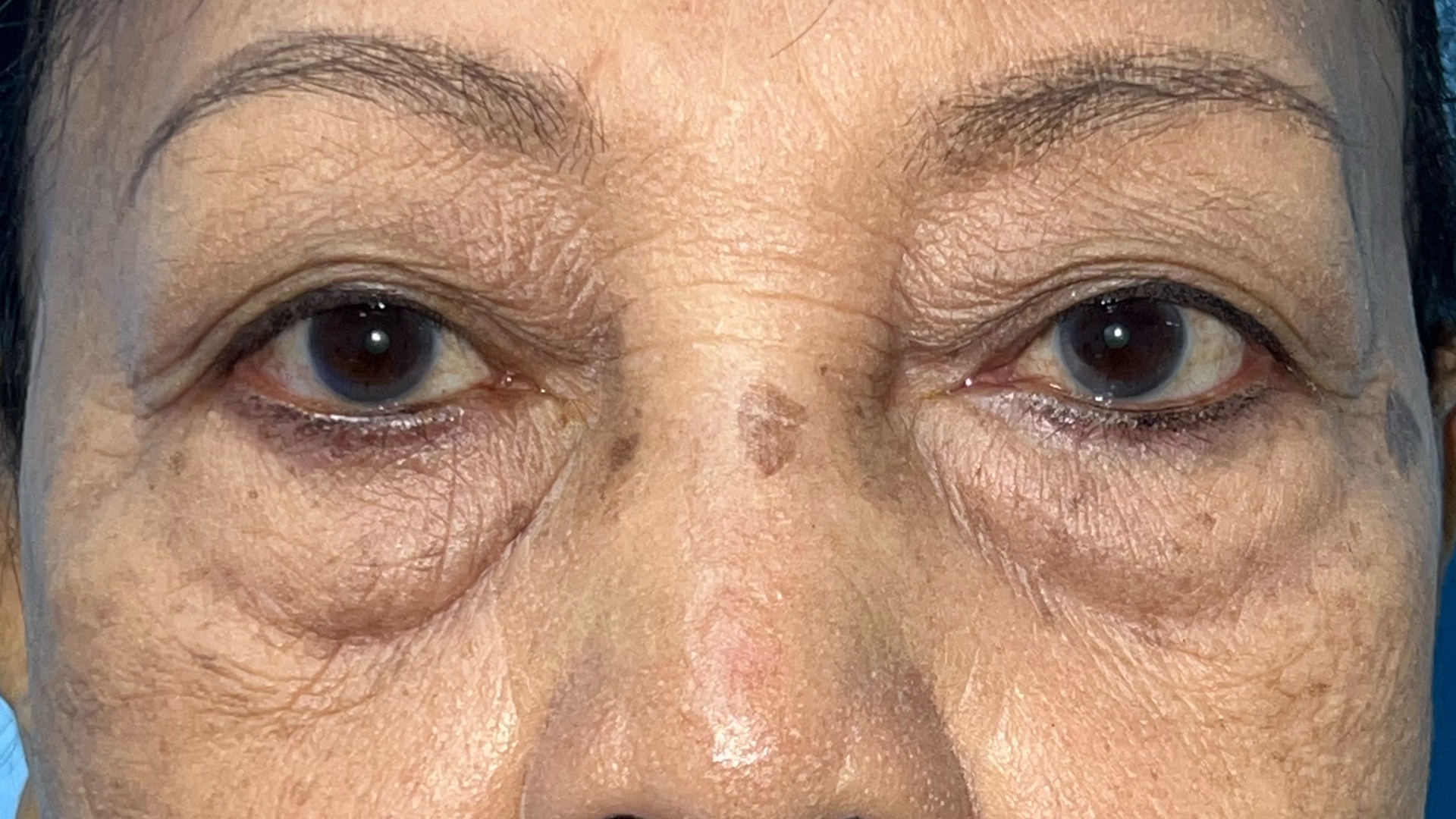
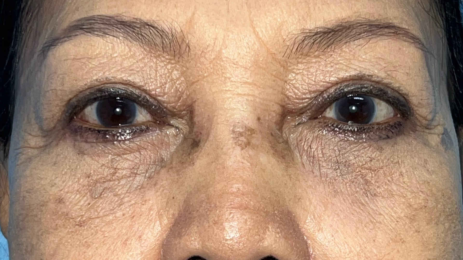
Prominent lower eyelid bags contributing to a persistently tired appearance. Lower blepharoplasty was used to address the lower eyelid contour.
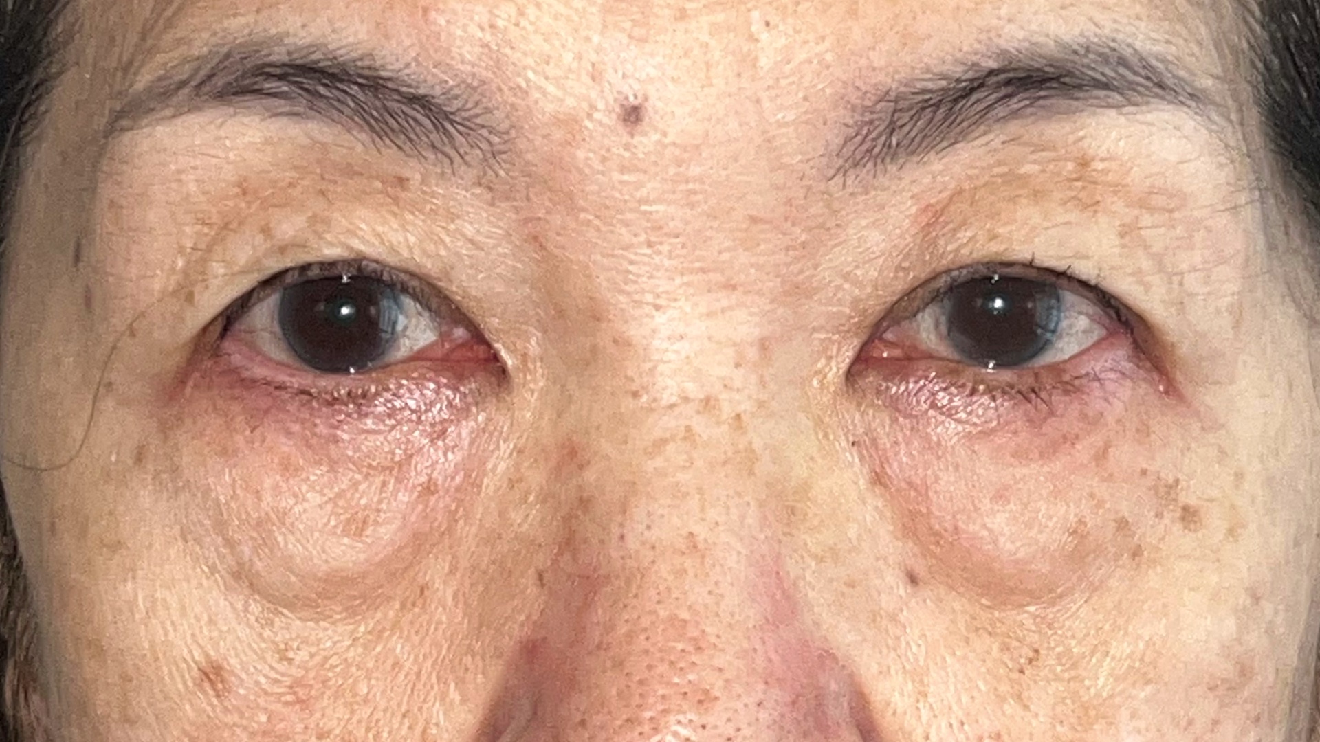
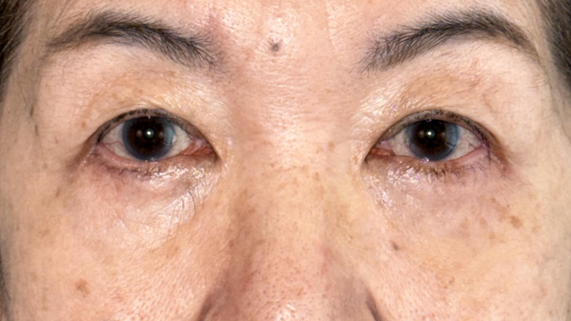
Before and 1-year postoperative photographs following lower blepharoplasty. Individual anatomy, treatment planning, and results vary between patients.
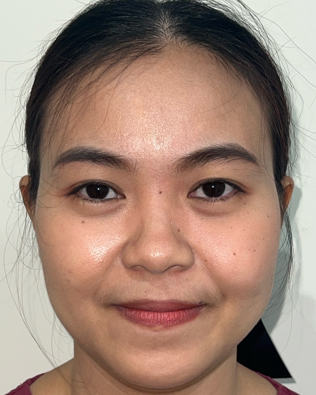
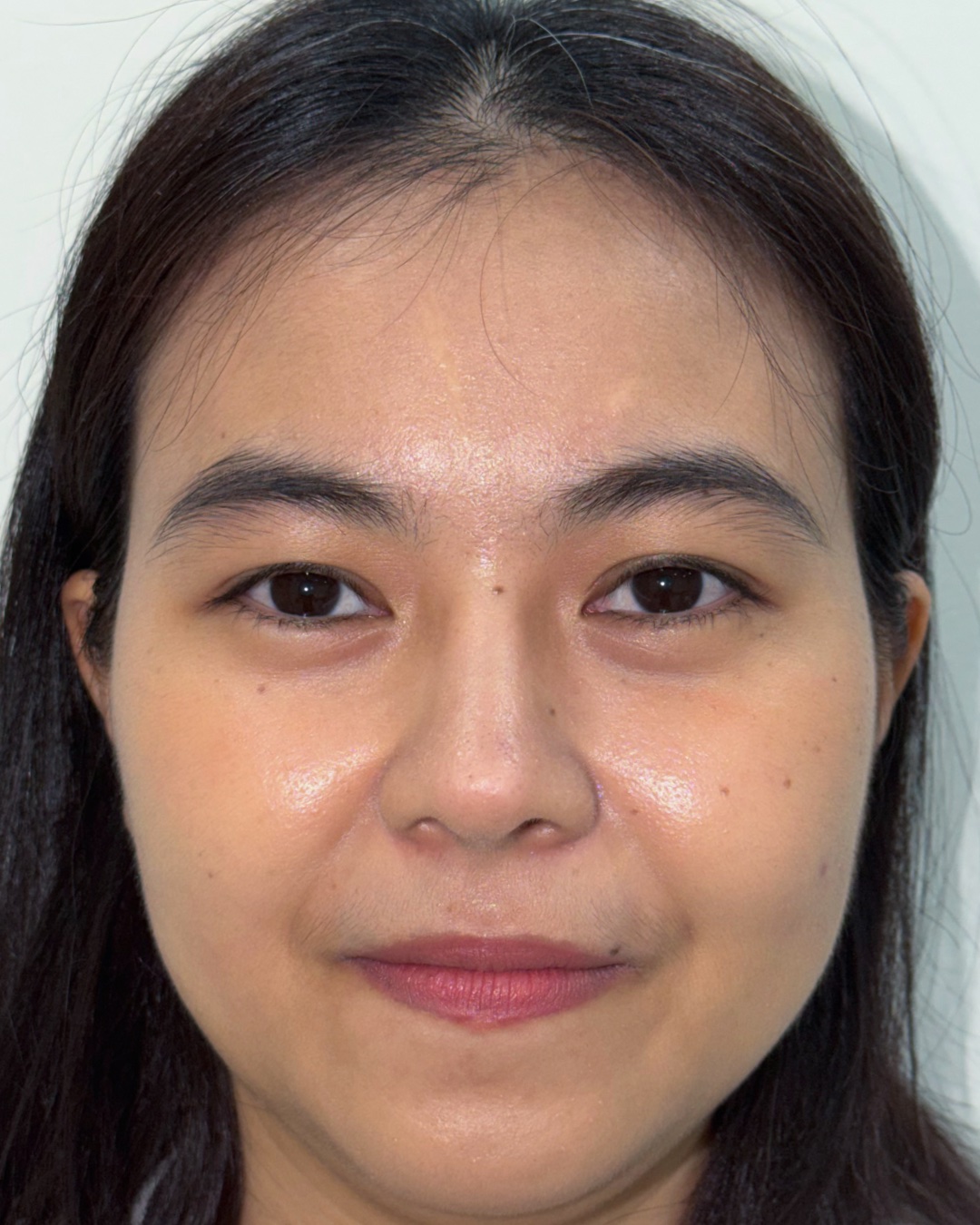
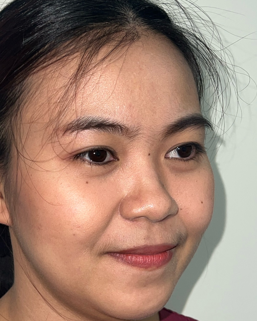
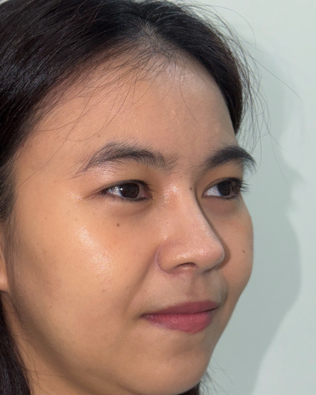
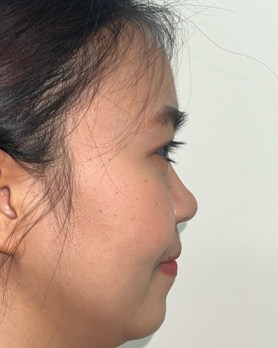
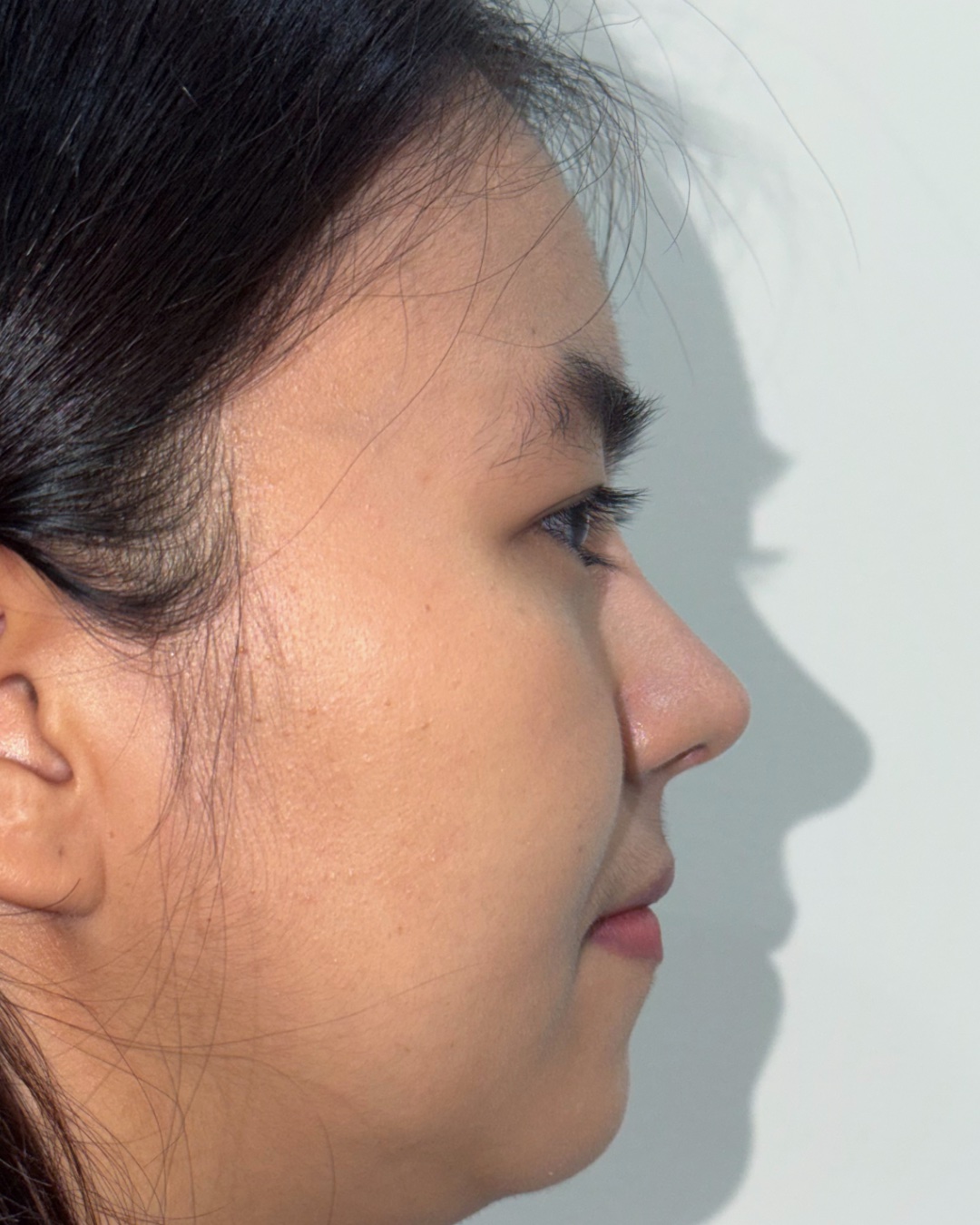
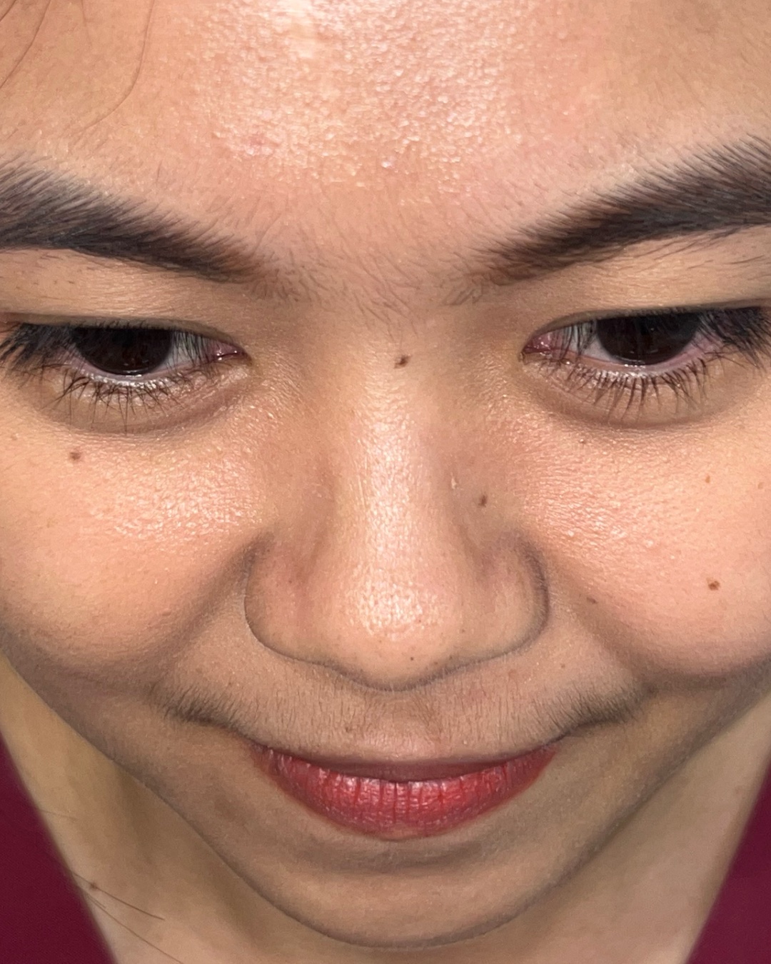
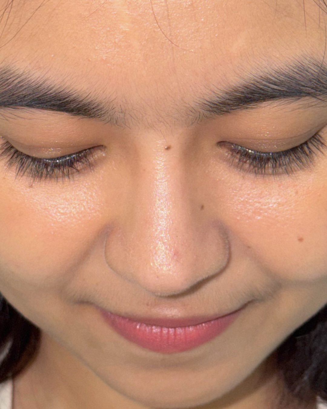
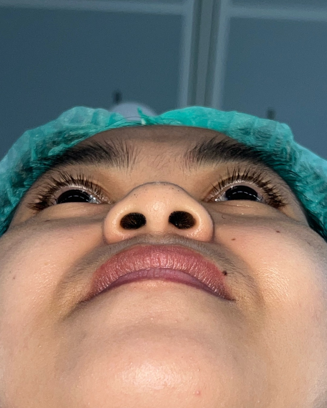
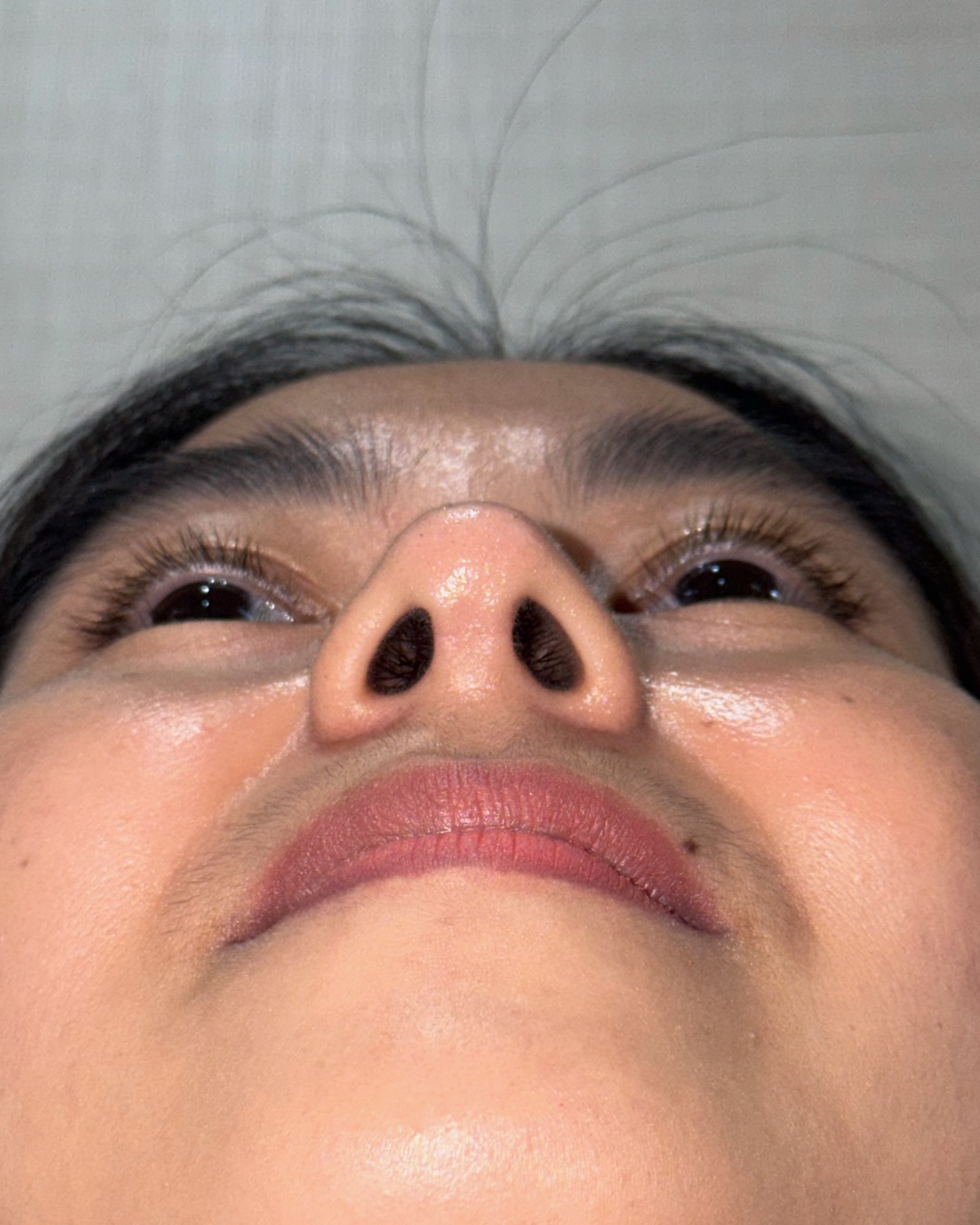
Before and 13-month follow-up photographs shown across matched front, oblique, profile, top-down and base views.
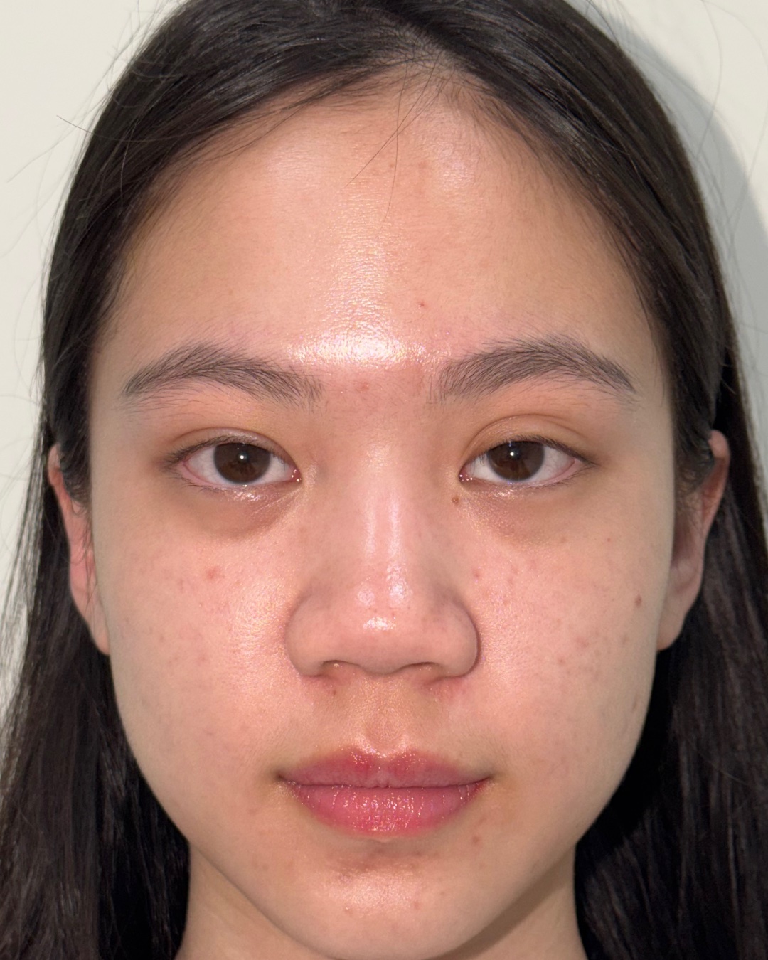
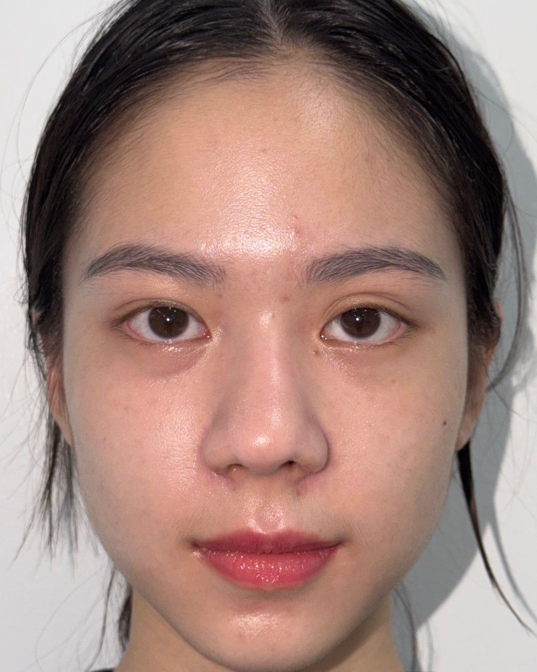
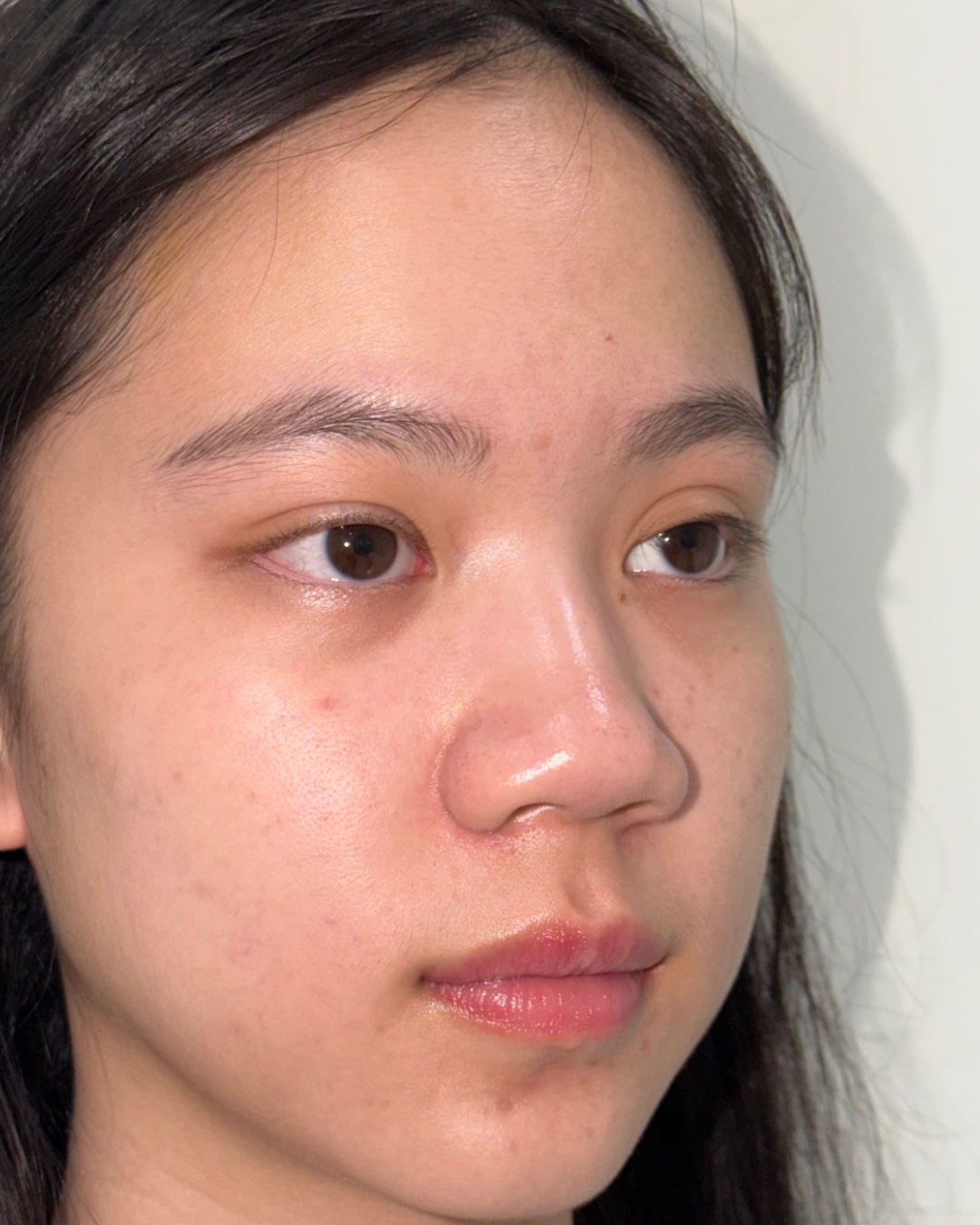
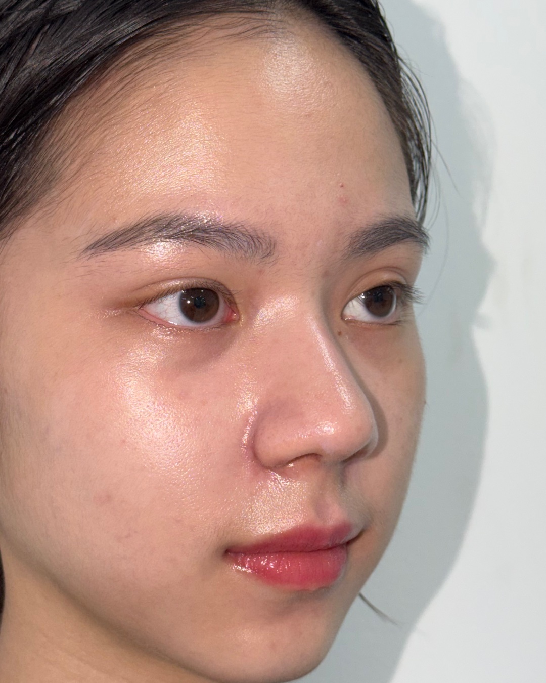
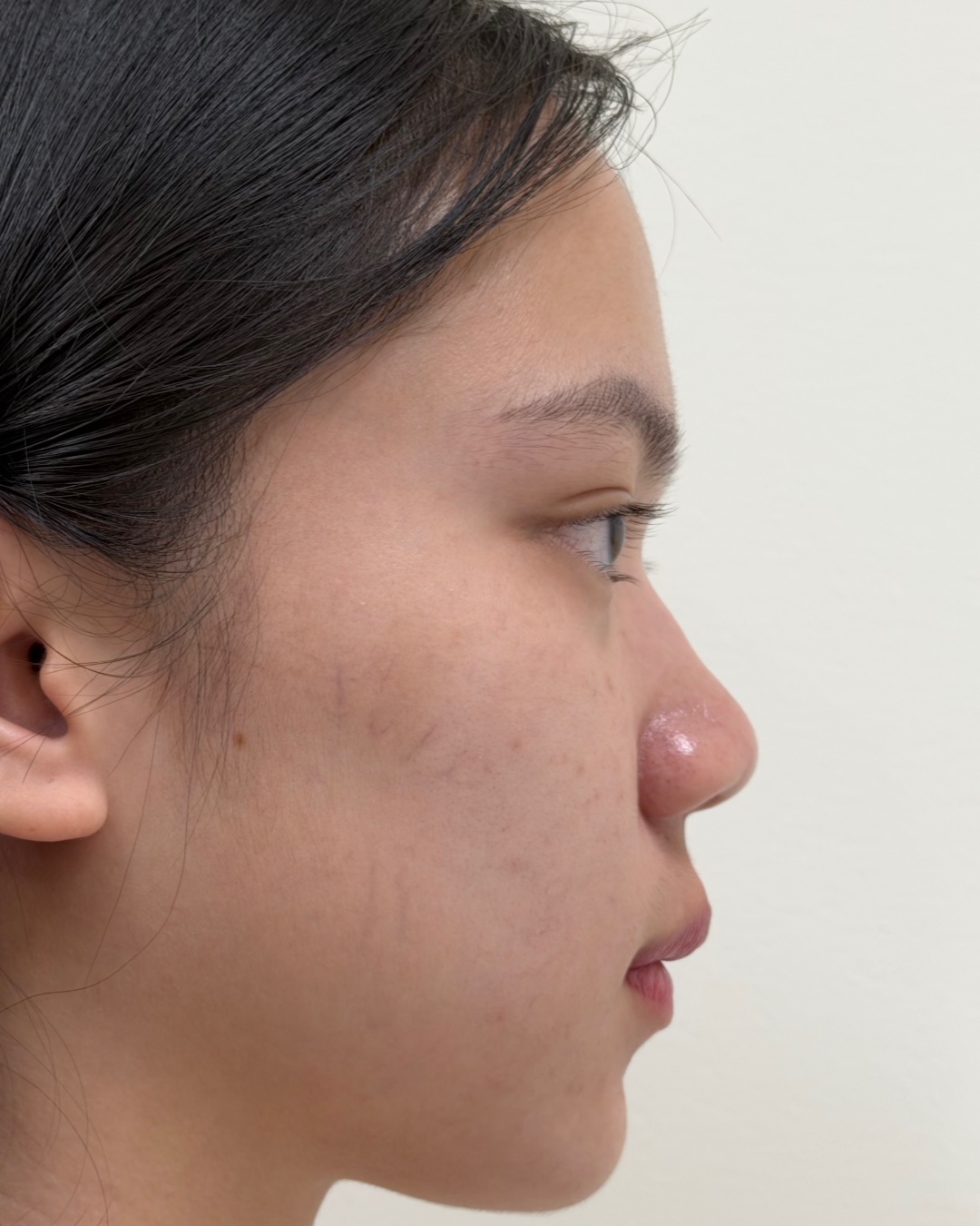
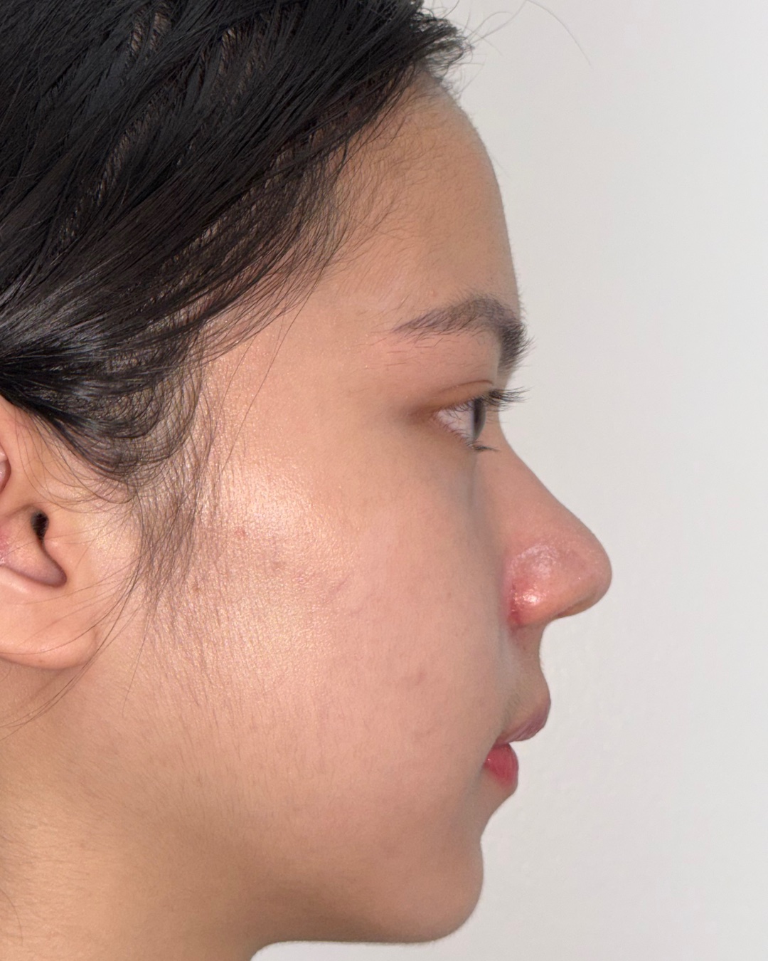
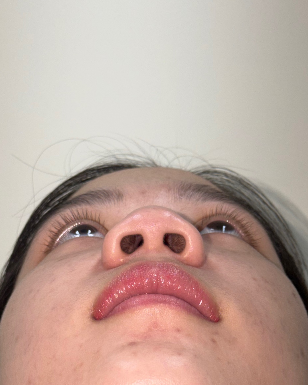
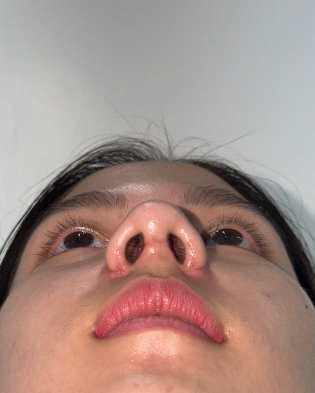
Before and 2-month follow-up photographs shown across matched front, oblique, profile and base views.
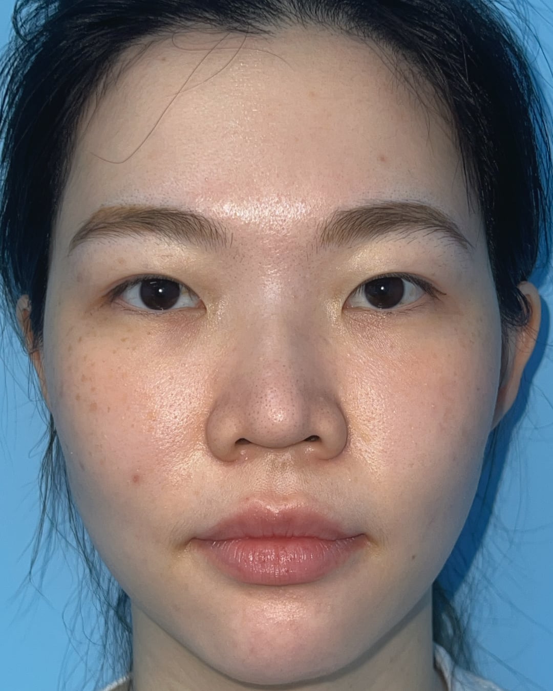
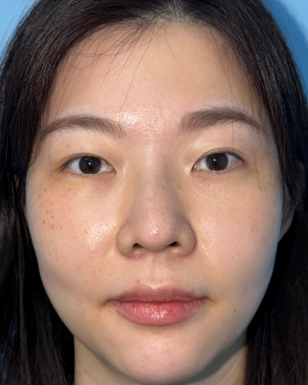
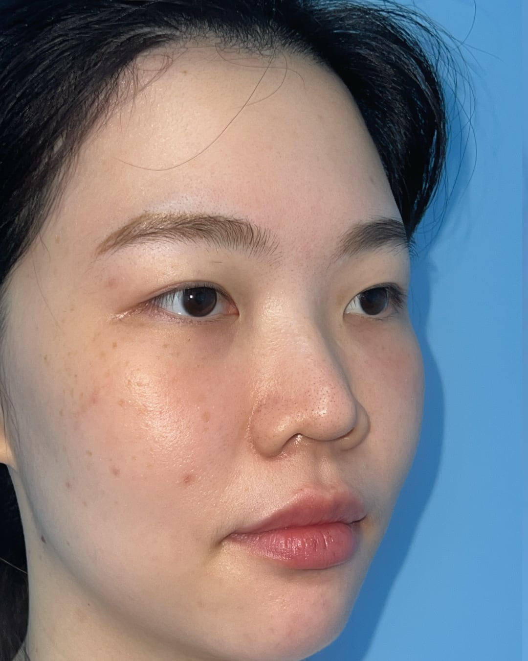
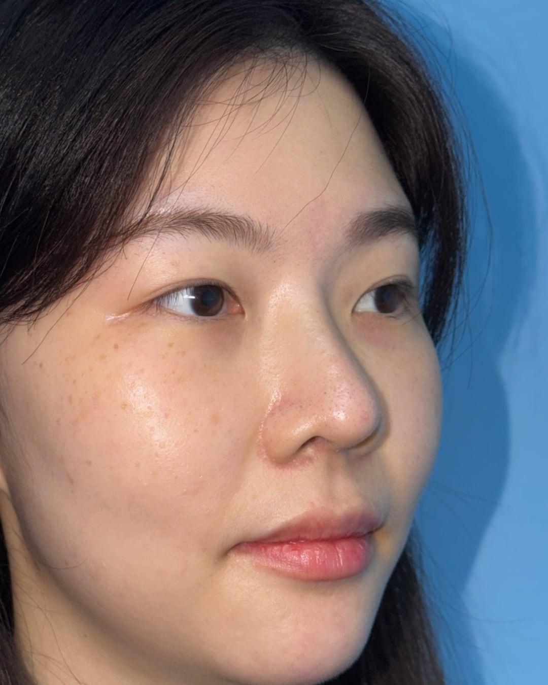
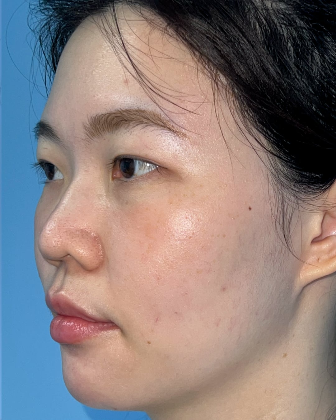
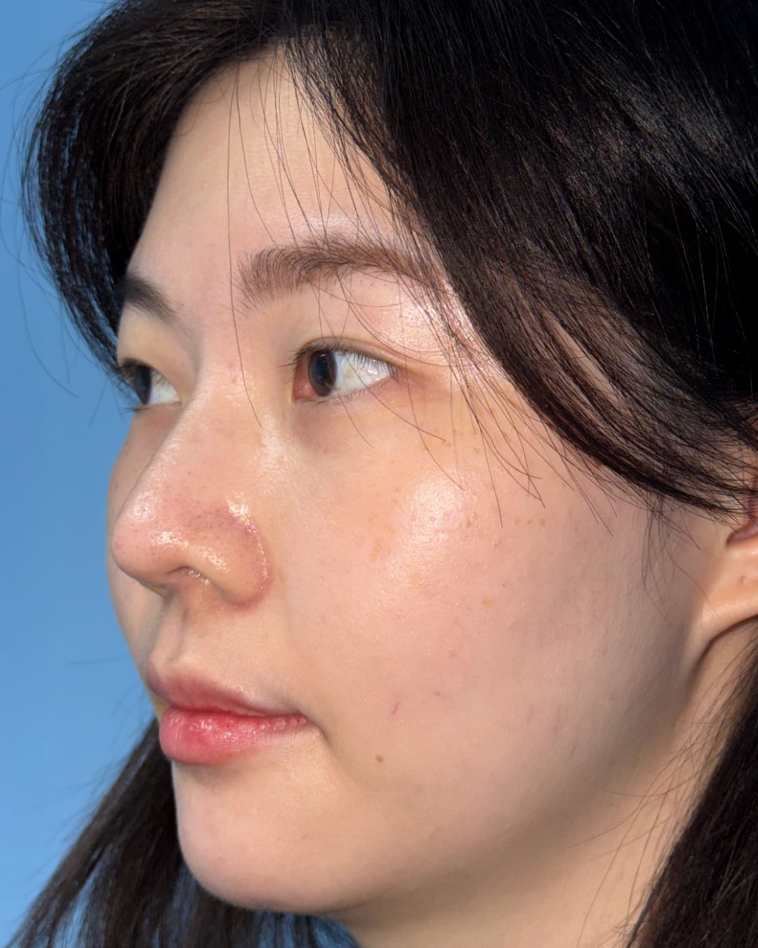
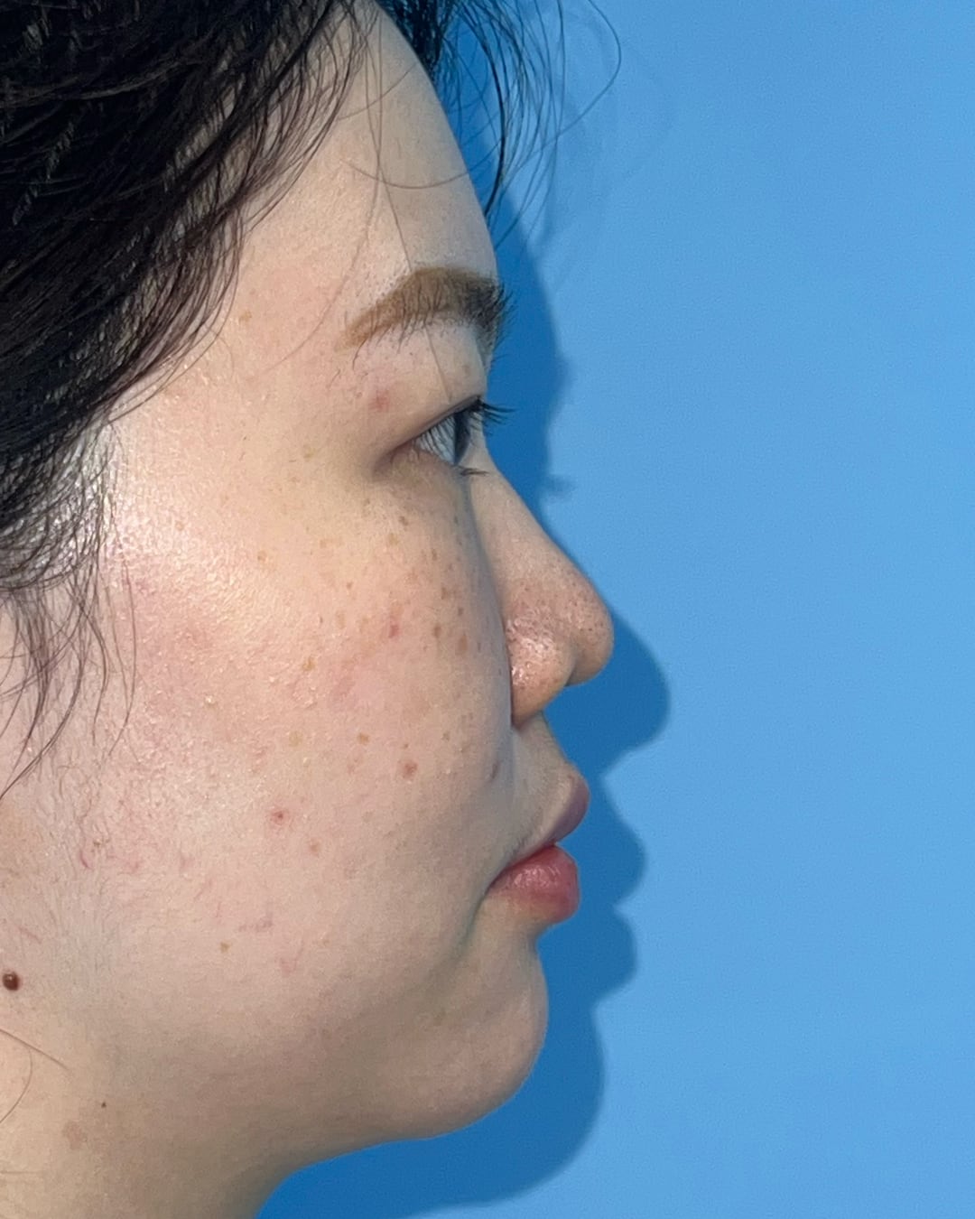
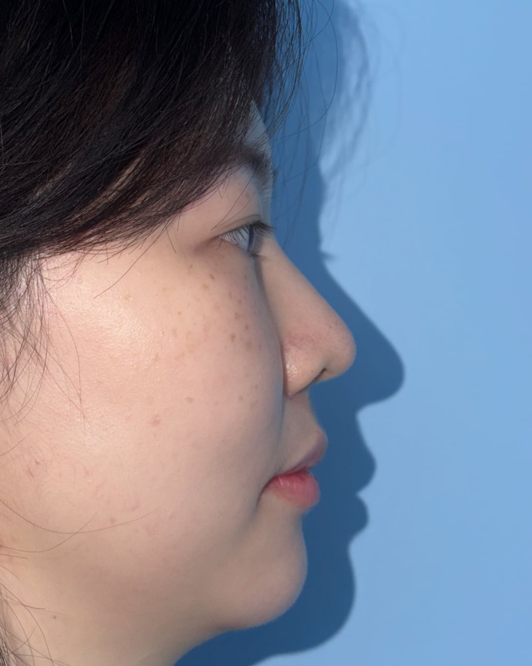
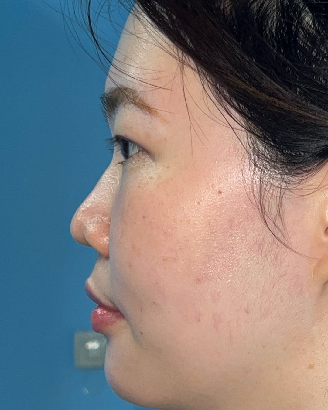
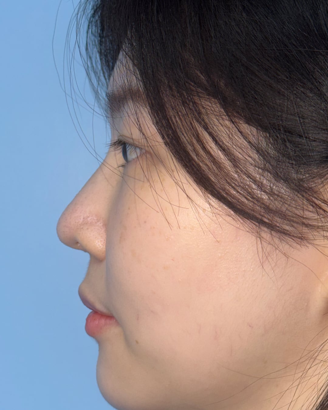
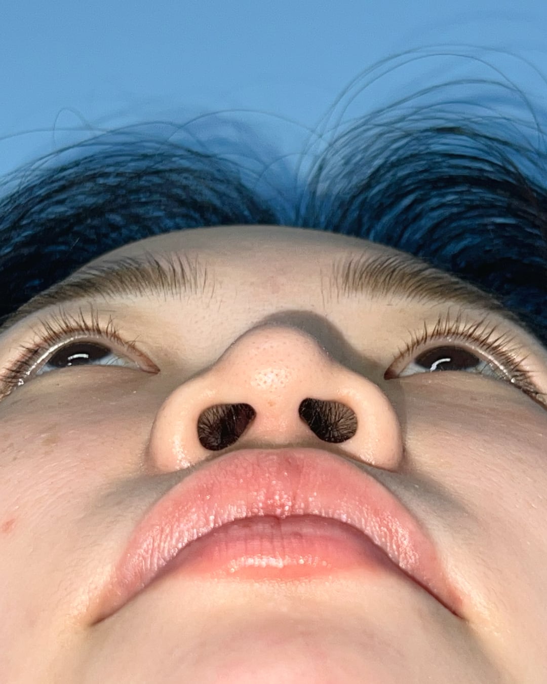
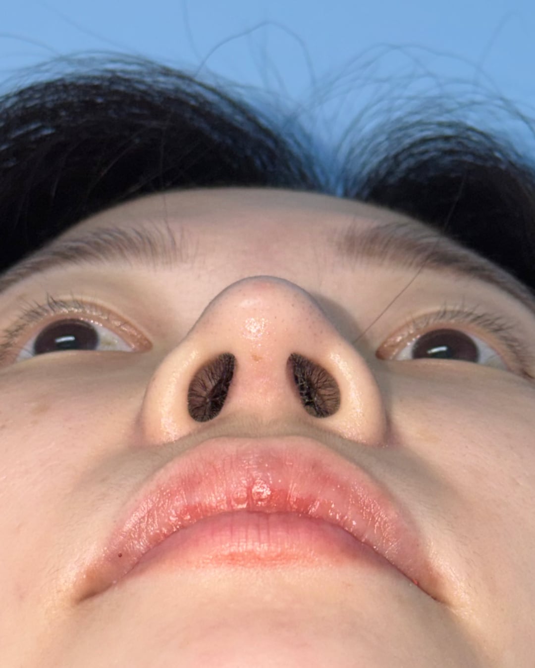
Before and 6-month follow-up photographs shown across matched AP, bilateral oblique, bilateral profile and base views.
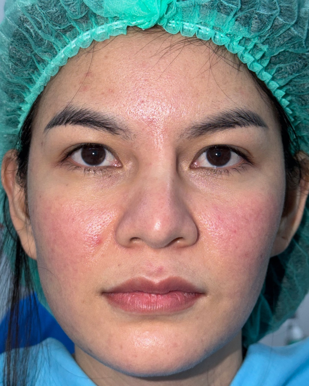
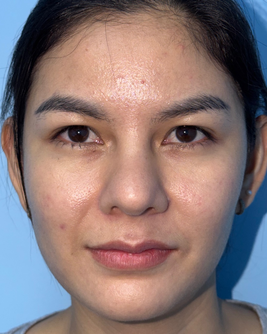
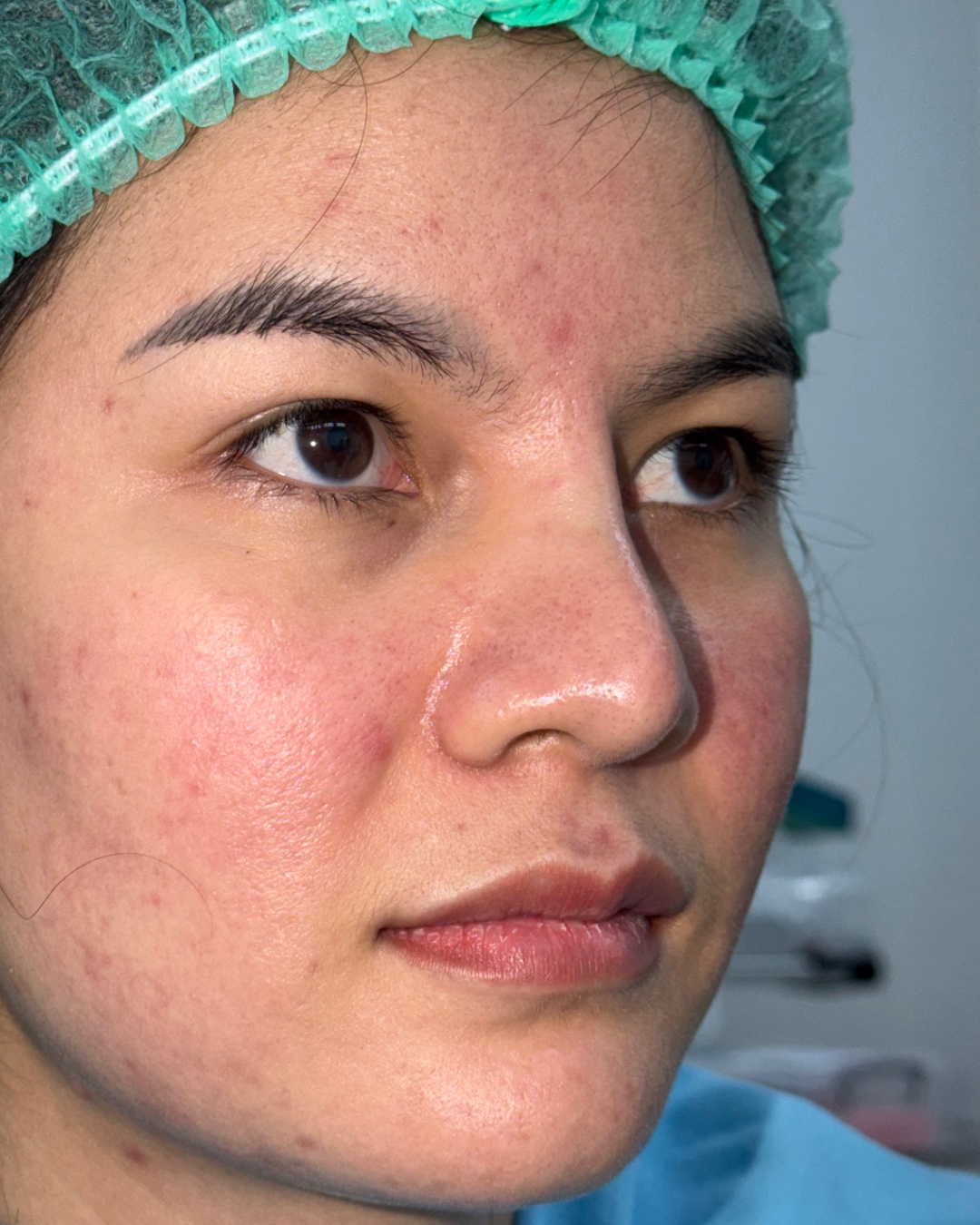
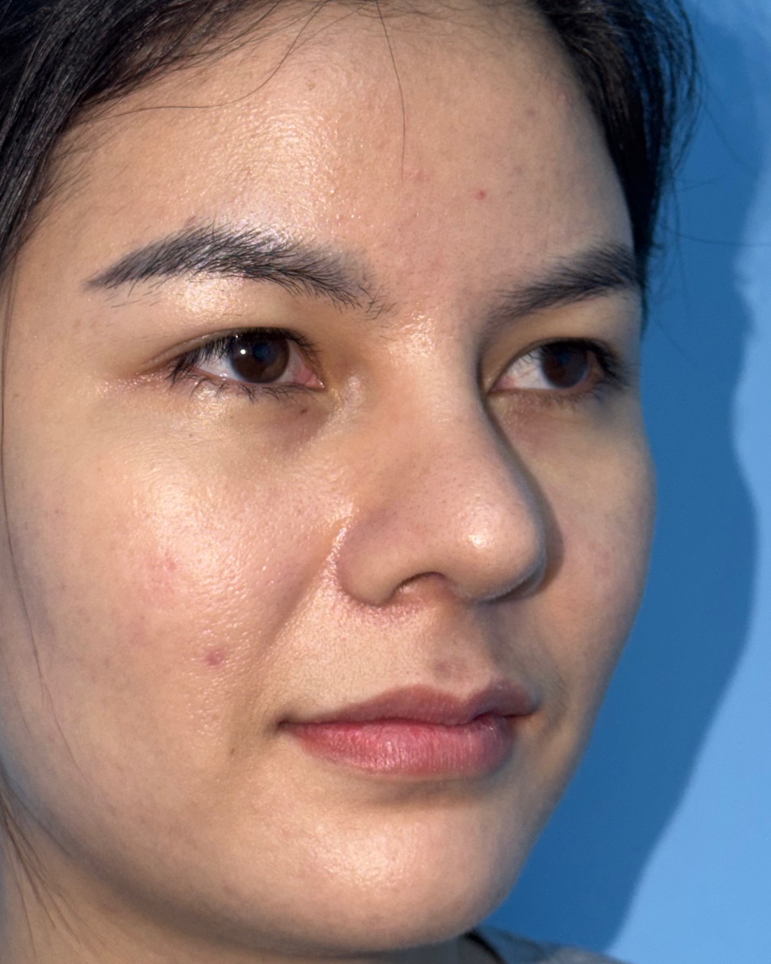
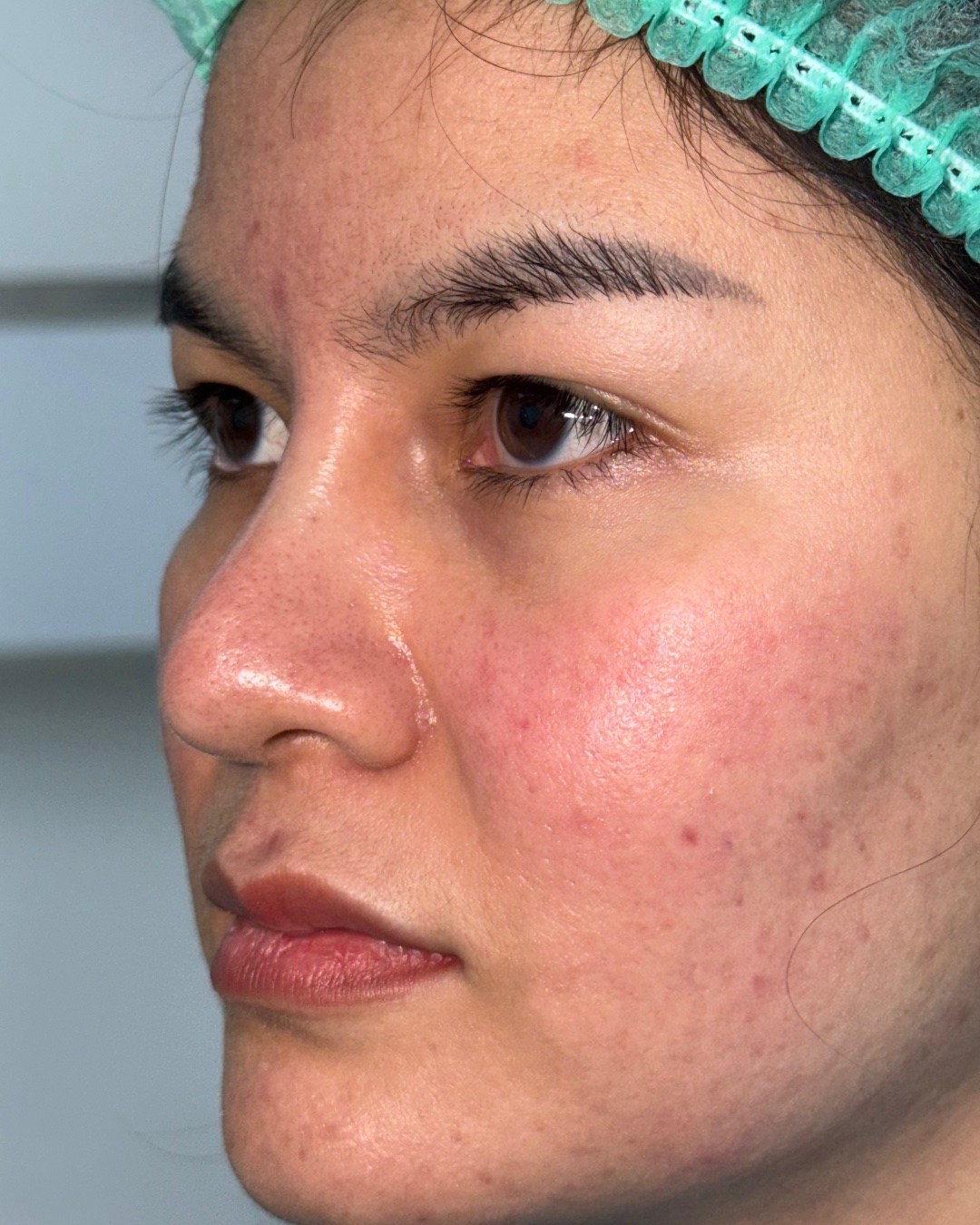
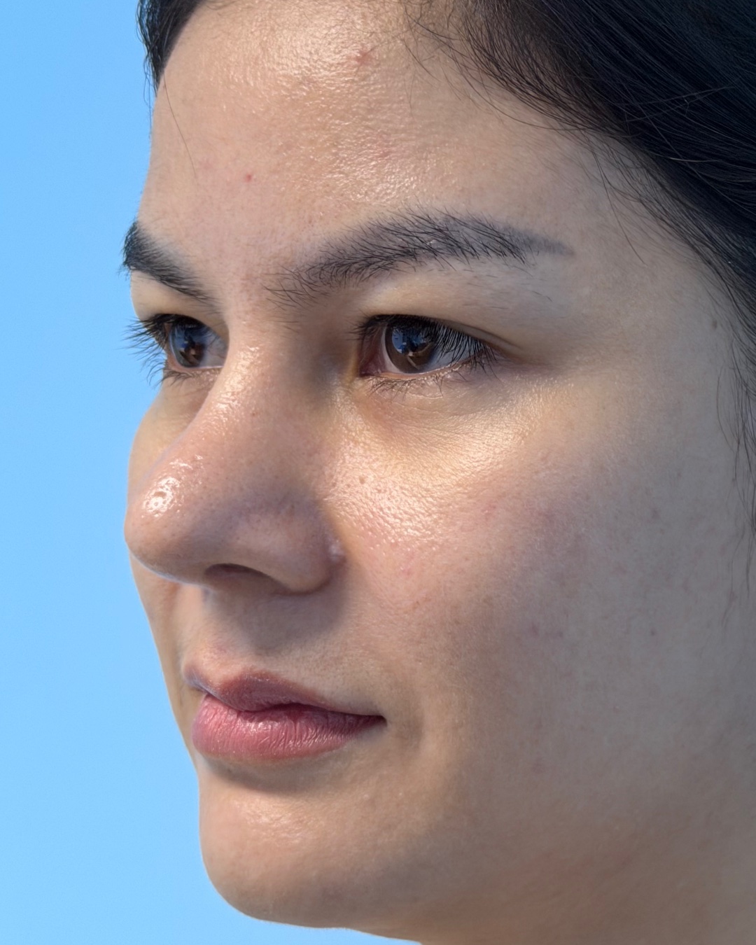
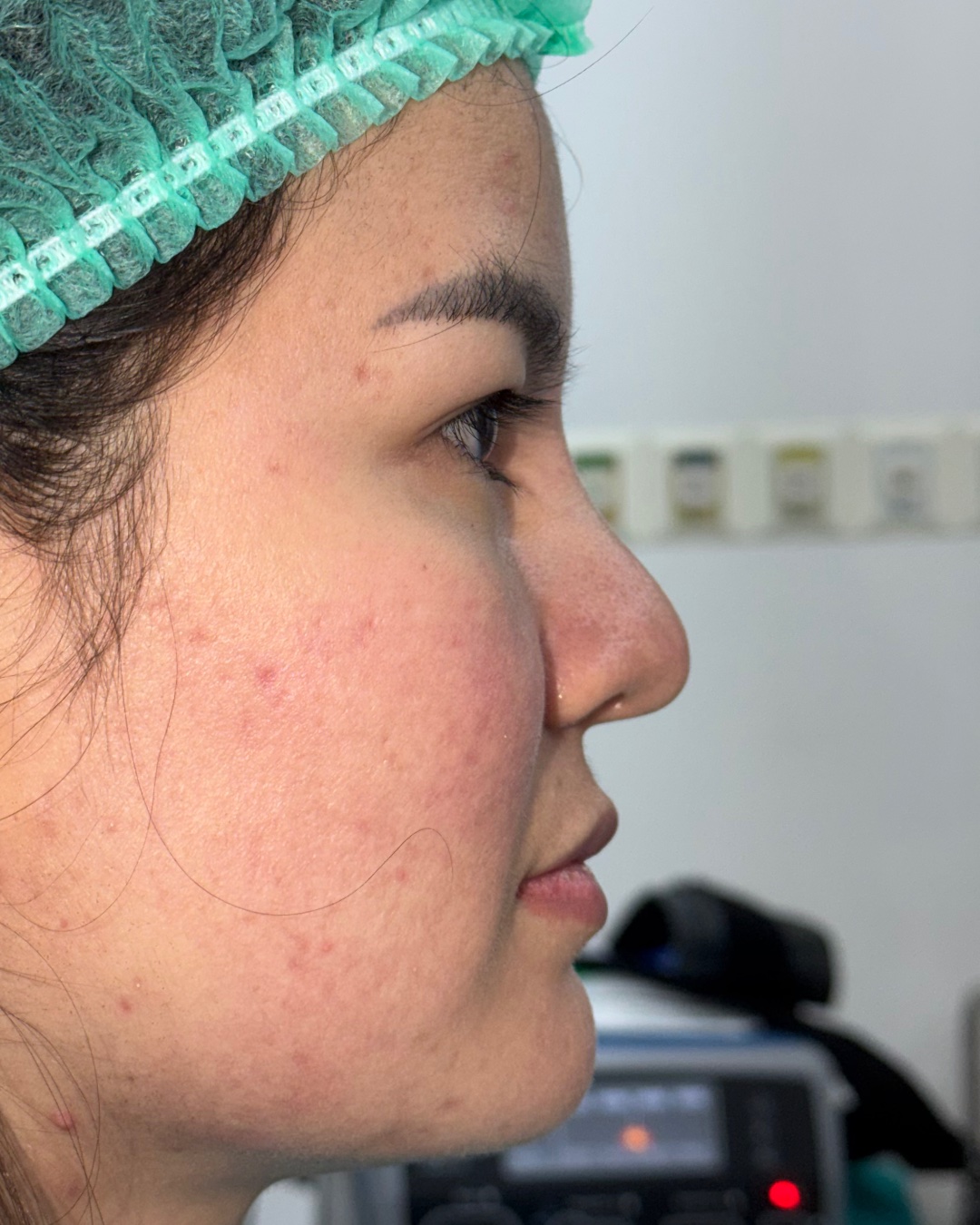
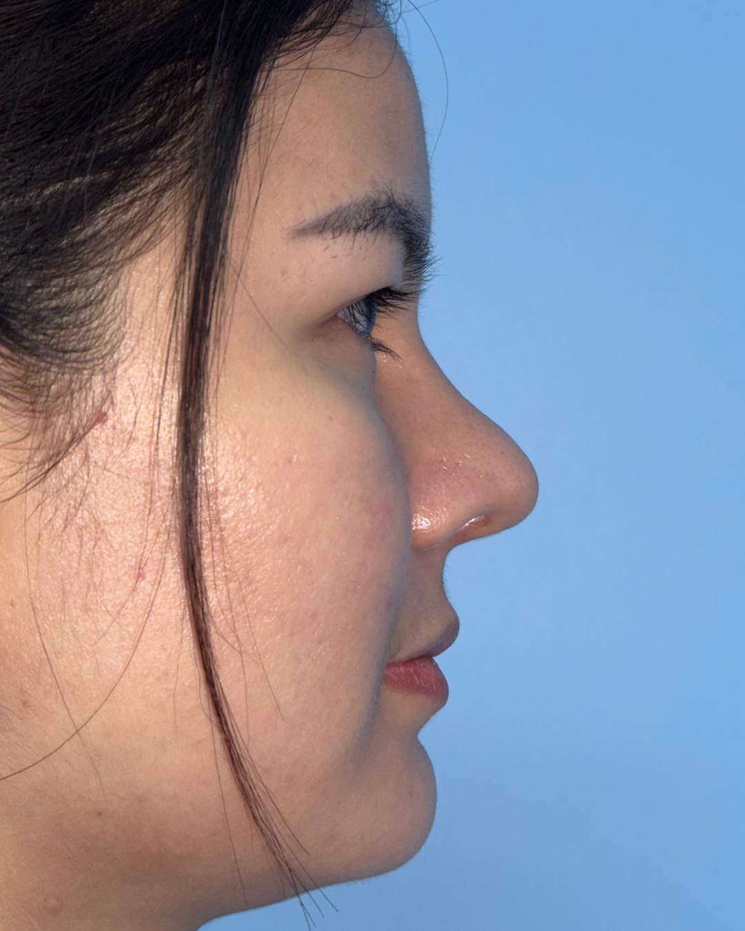
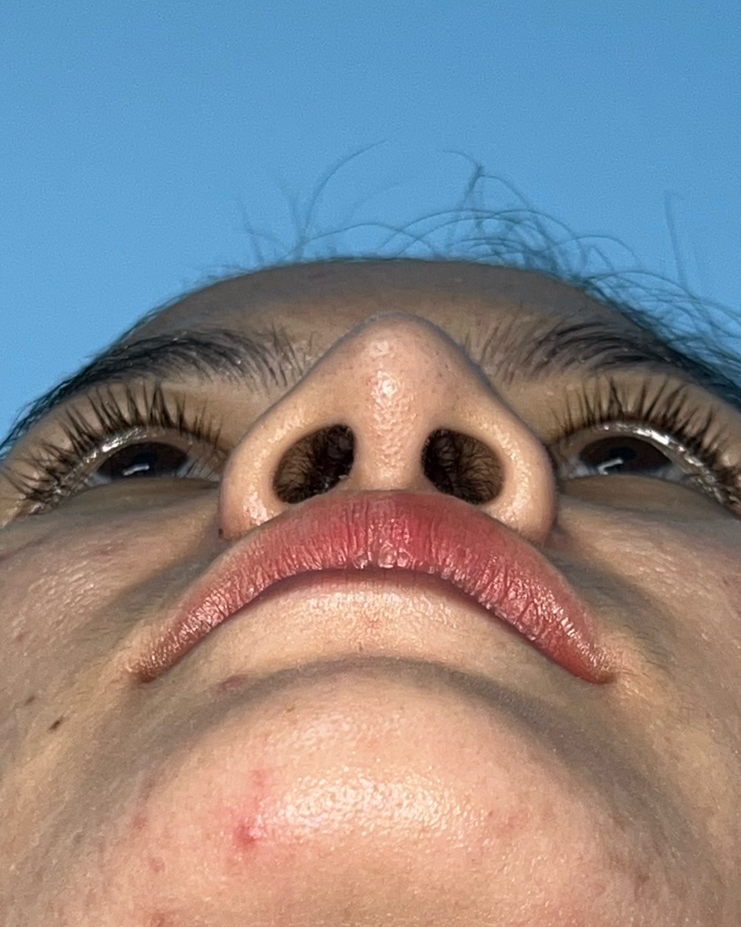
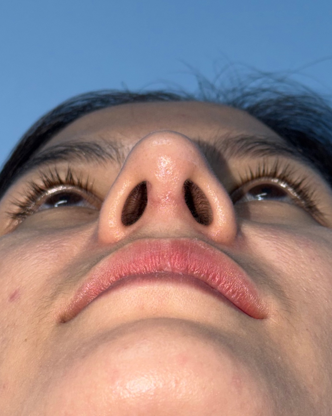
Before and 4-month follow-up photographs shown across matched front, bilateral oblique, right profile and base views.
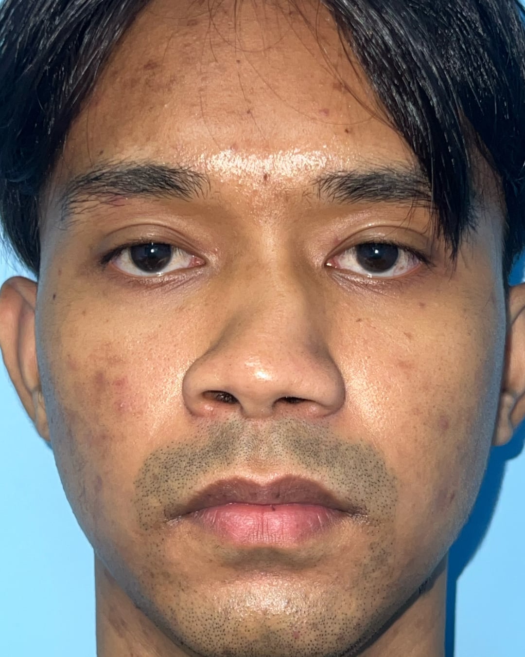
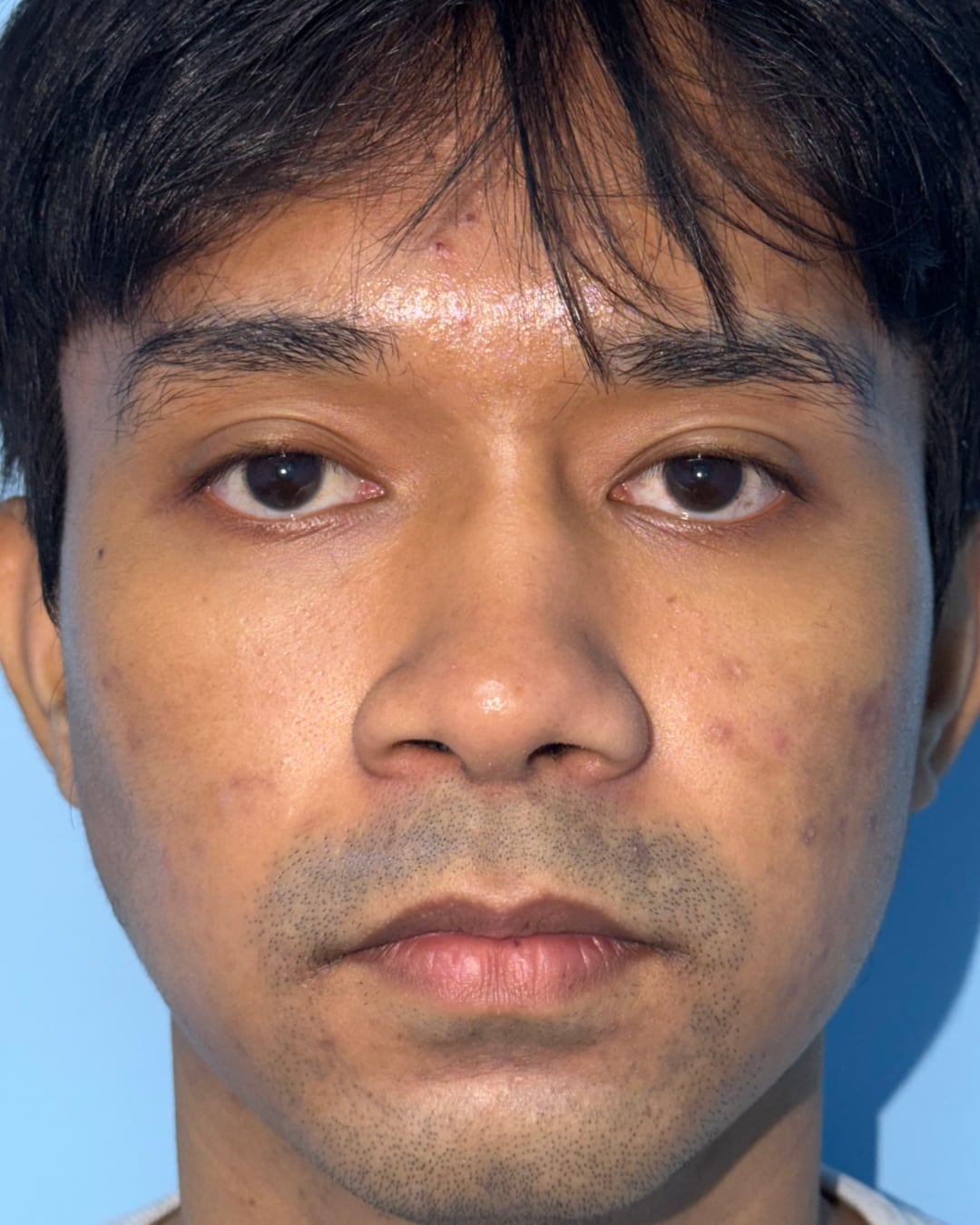
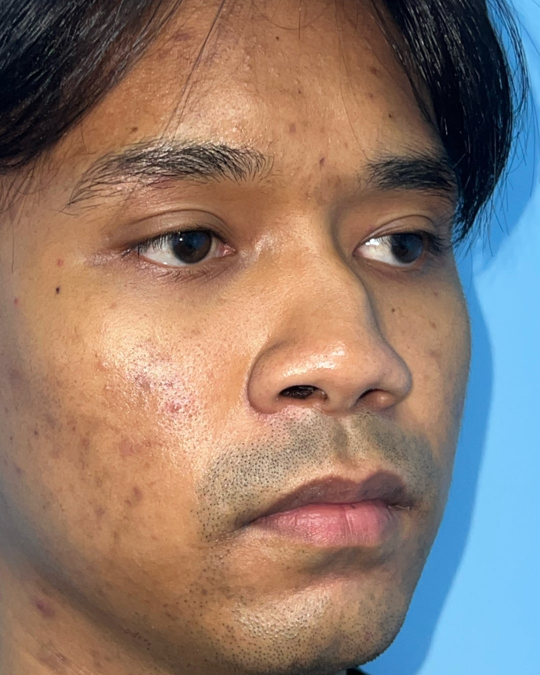
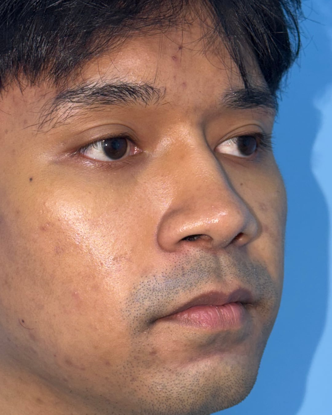
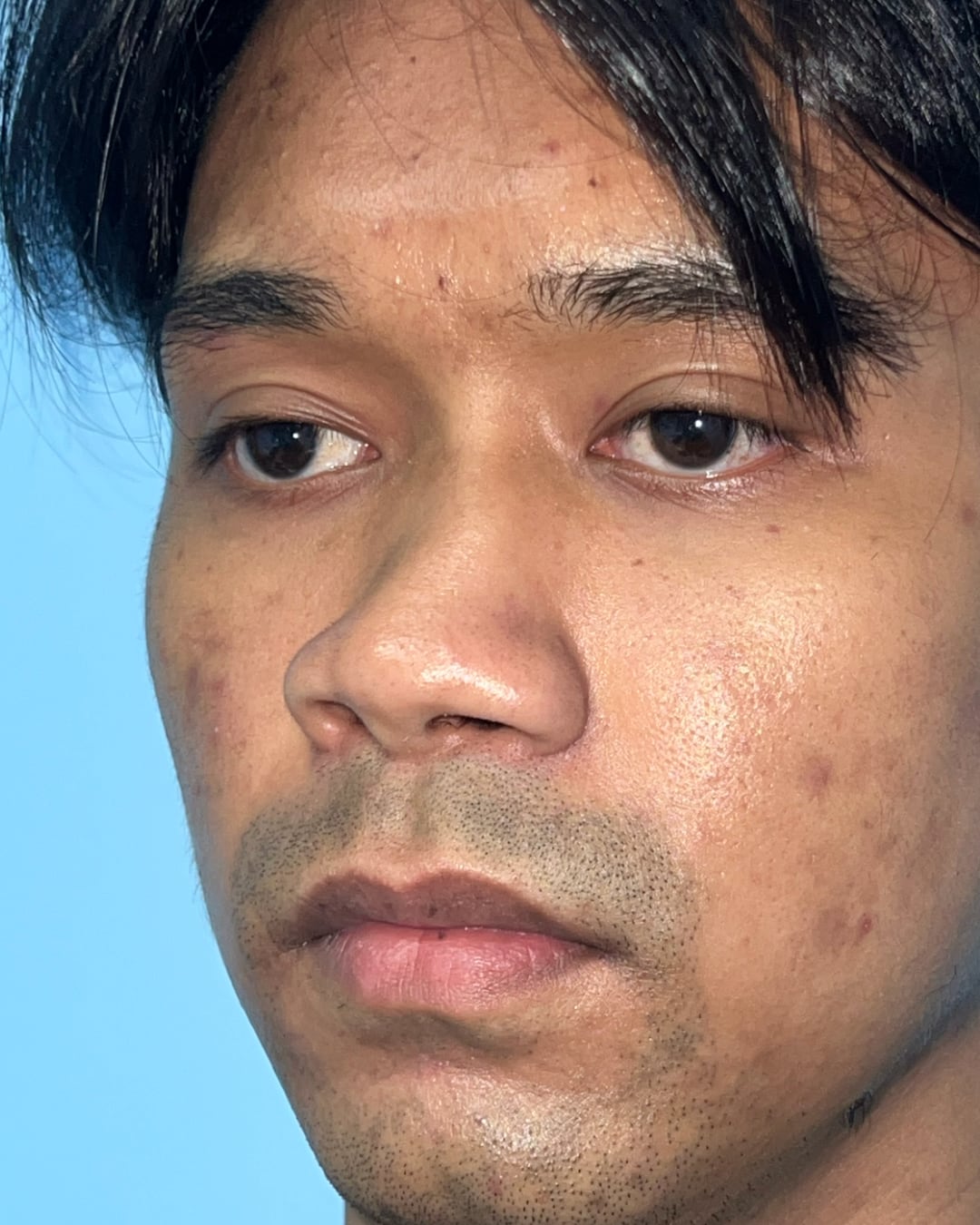
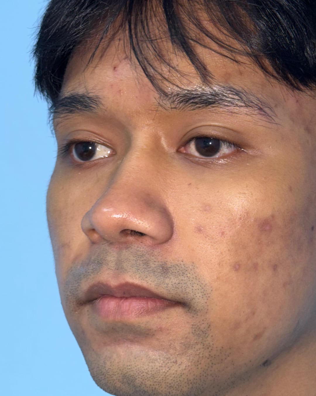
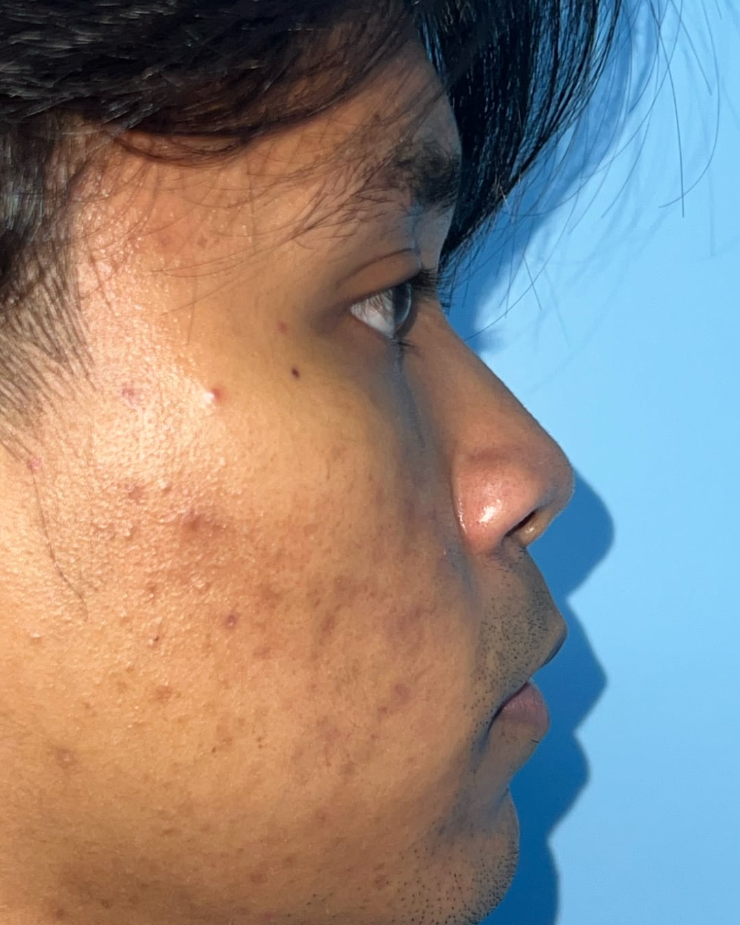
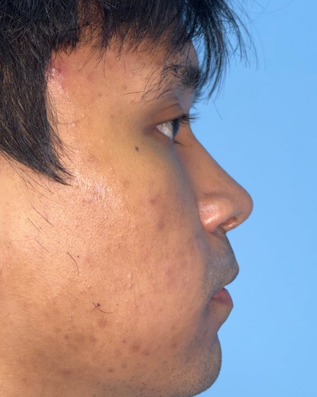
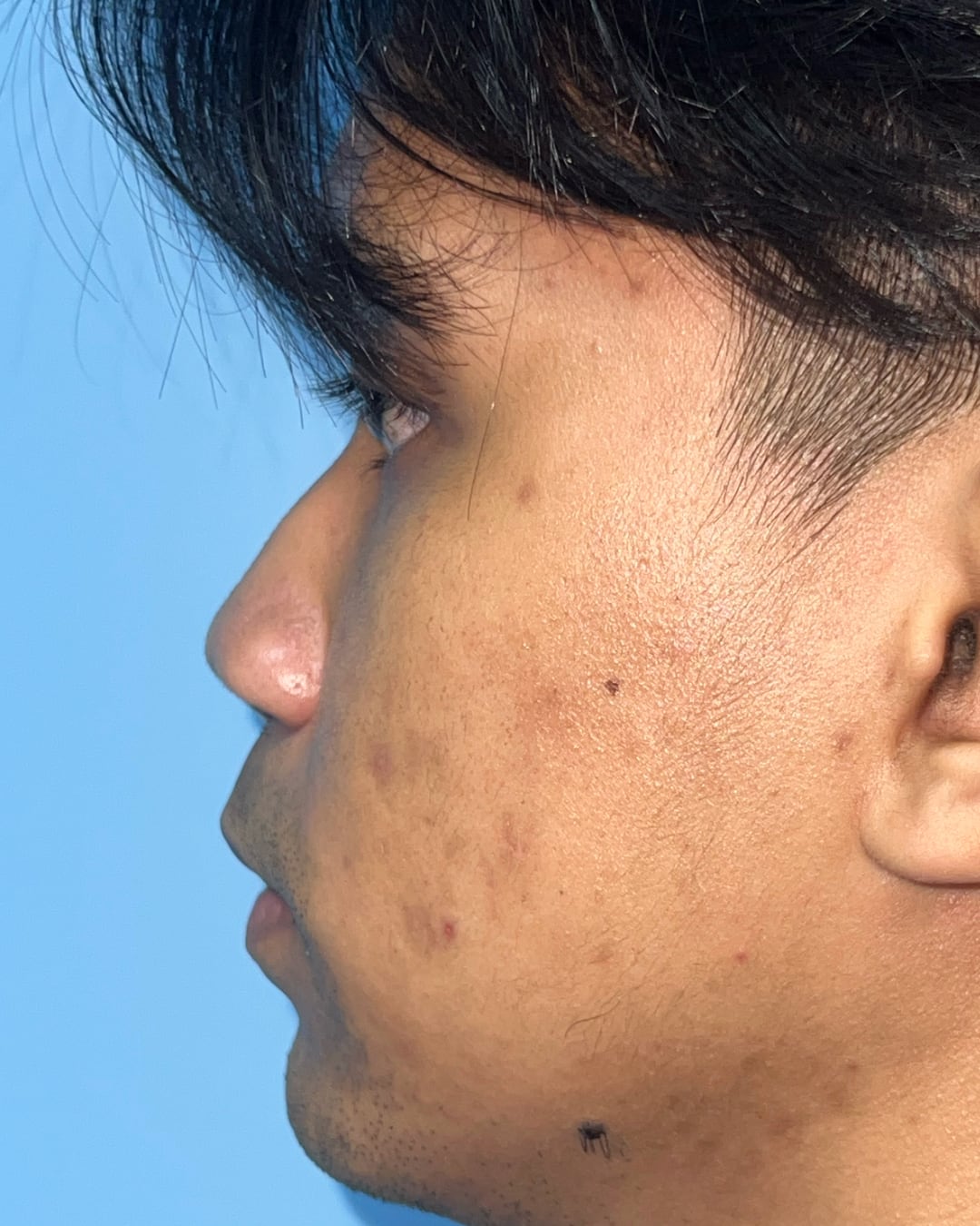
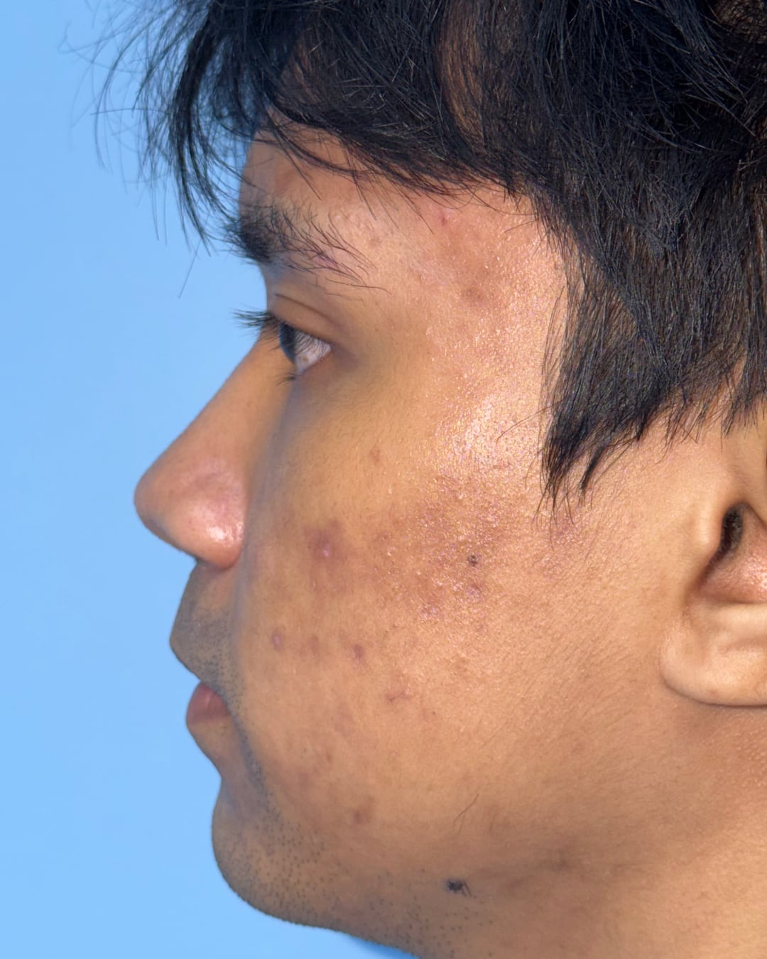
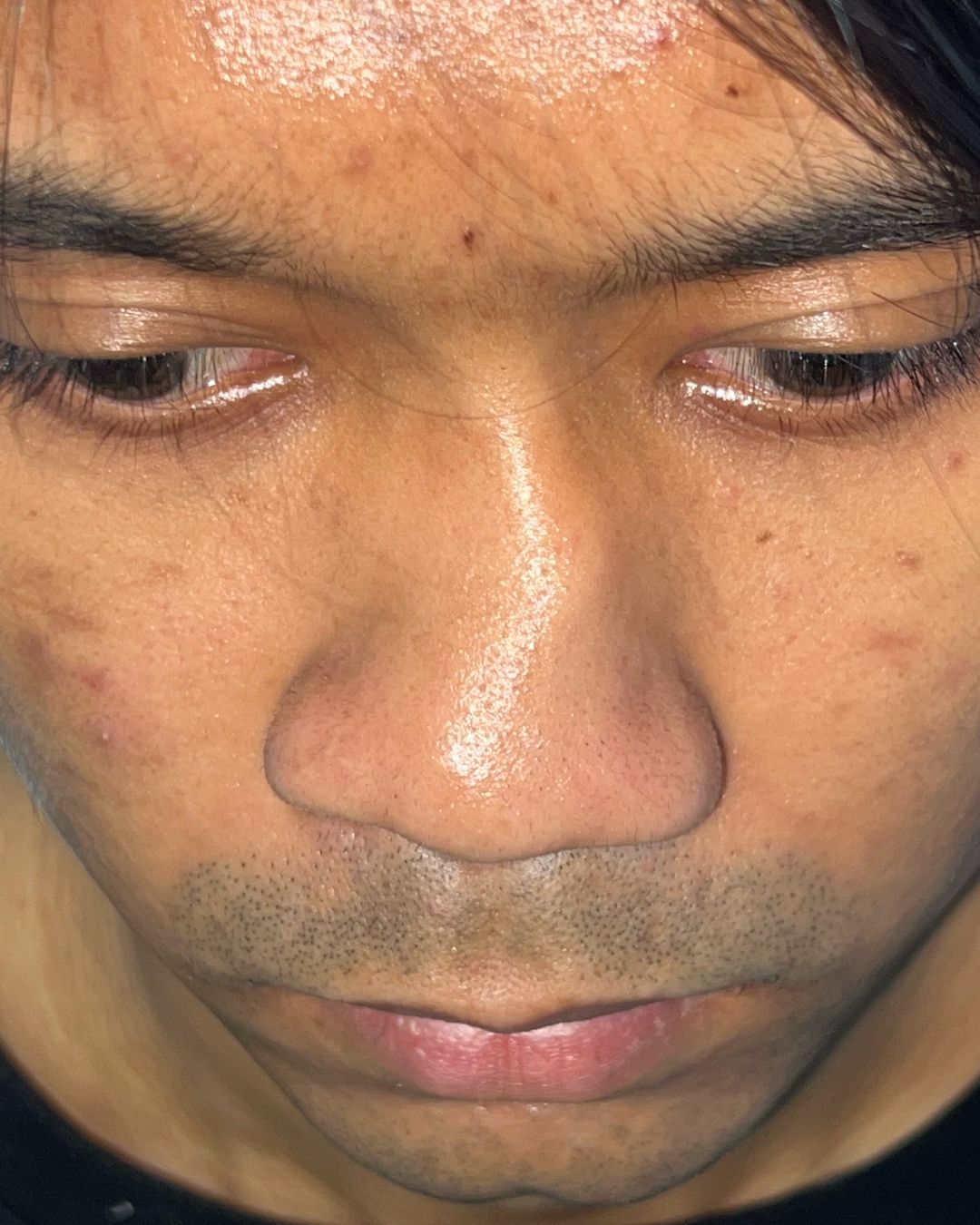
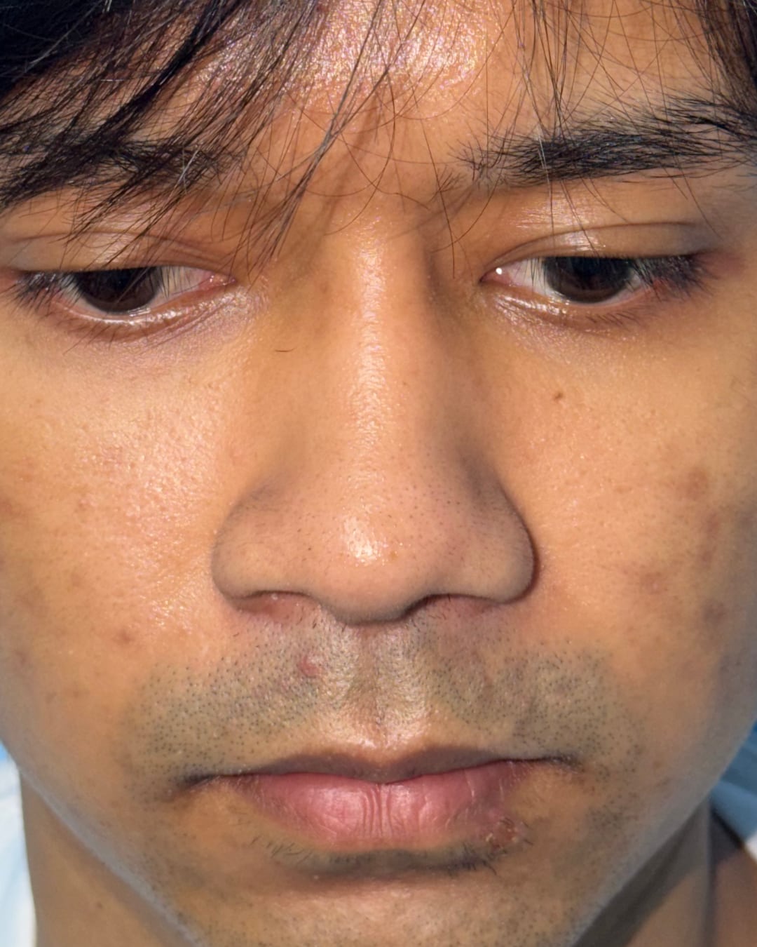
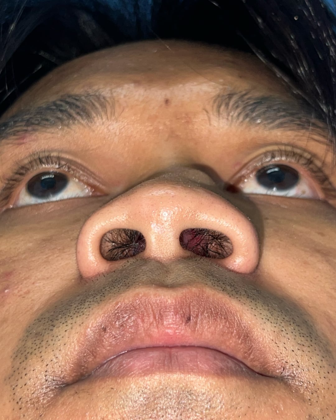
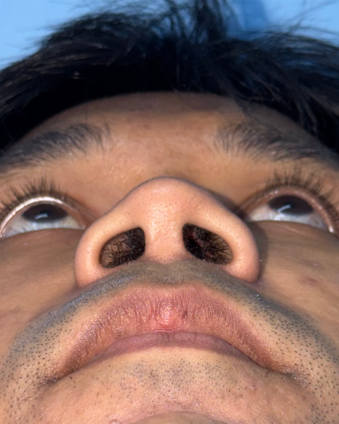
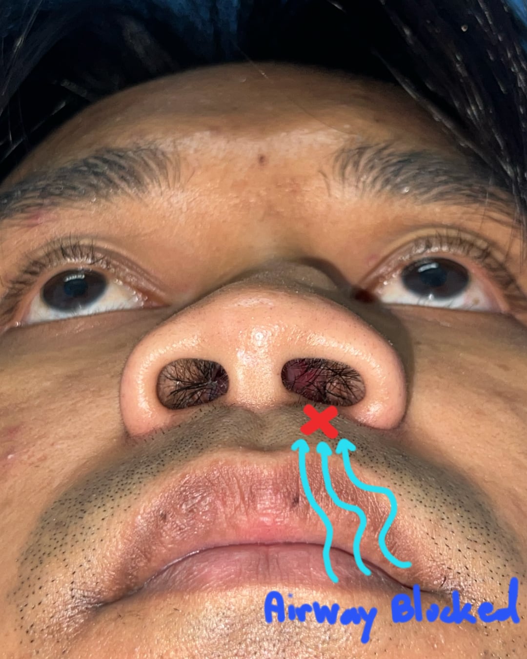
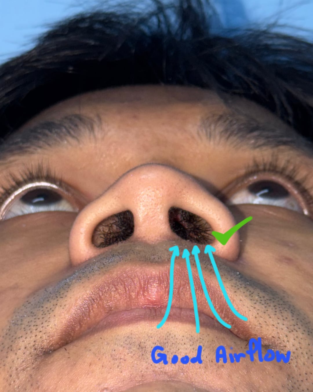
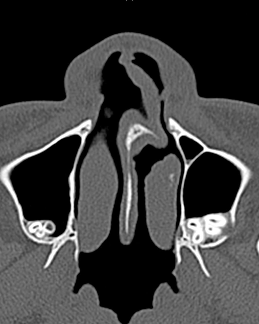
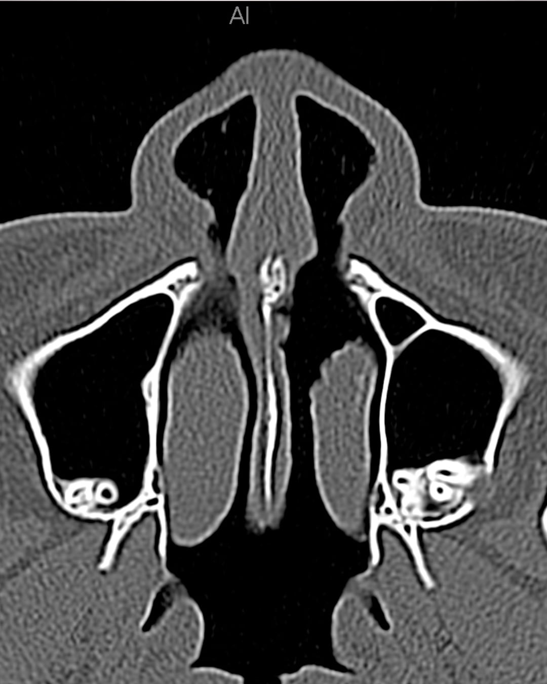
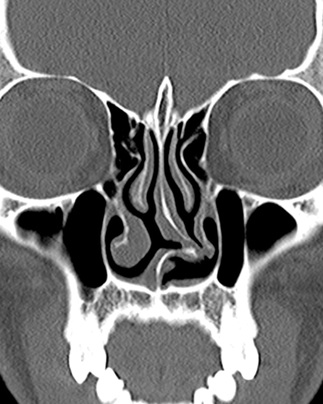
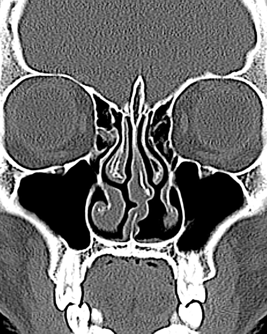
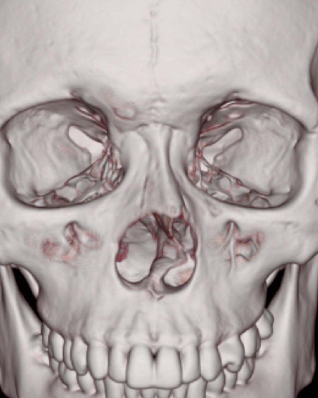
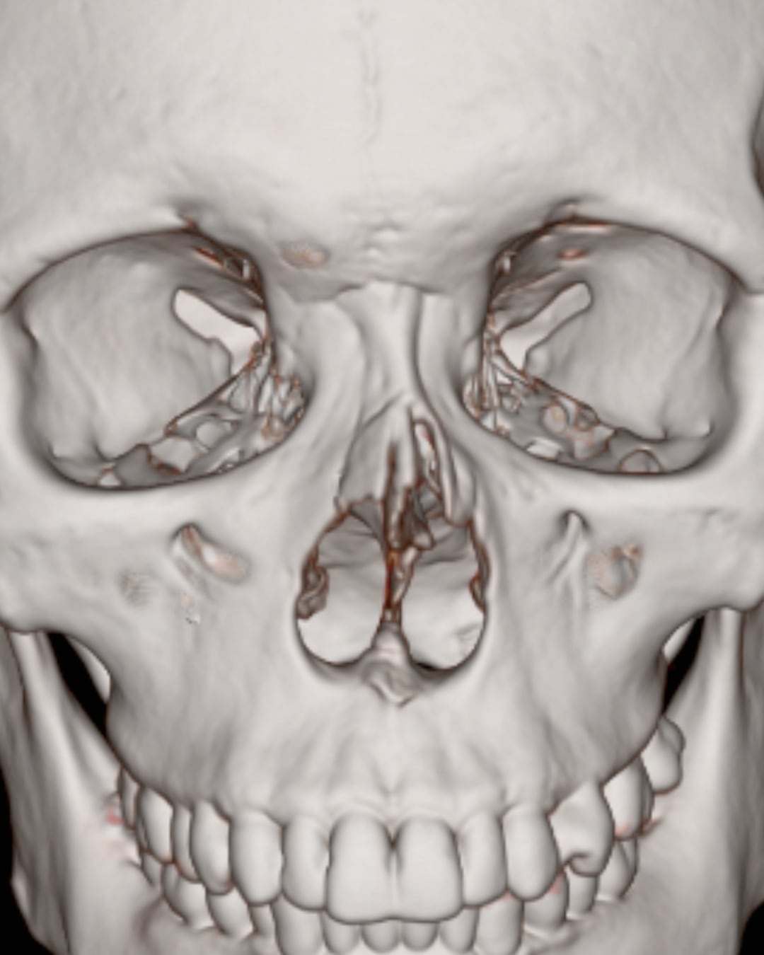
Severe congenital nasal deviation treated with an open rhinoplasty approach and costal cartilage grafting. One-year follow-up includes matched clinical photographs, functional airflow views, and CT imaging.
Patient photographs are presented for educational purposes with appropriate permission. Individual anatomy, indications, healing and outcomes vary. A consultation and physical examination are required for an individual assessment.
Before and after photographs can help explain surgical possibilities, but treatment should be planned around your own anatomy, function, goals and indications.